 Entrance
Which algae do not have flagellar stages? Class Flagellates
Monadic vegetative cells and monadic stages in the life cycle (zoospores and gametes) of algae are equipped with flagella - long and rather thick cell outgrowths, externally covered with plasmalemma. Their number, length, morphology, place of attachment, and pattern of movement are quite diverse in algae, but are constant within related groups.
The flagella may be attached at the anterior end of the cell (apical) or may be slightly moved to the side (subapical); they can be attached to the side of the cell (laterally) and on the ventral side of the cell (ventrally). Flagella identical in morphology are called isomorphic, if they differ - heteromorphic. Isocont- these are flagella of the same length, heterokontnye– different lengths.
Flagella have a single structure plan. One can distinguish the free part (undulipodium), transition zone, and basal body (kinetosome). Different parts of the flagellum differ in the number and arrangement of microtubules, which form the skeleton (Fig. 2).
Rice. 2. Scheme of the structure of algae flagella (according to: L.L. Velikanov et al., 1981): 1 – longitudinal section of the flagella; 2, 3 – transverse section through the tip of the flagellum; 4 – transverse section through the undulipodium; 5 – transition zone; 6 – cross section through the base of the flagellum – kinetosome
Undulipodium(translated from Latin as “wavepod”) is capable of making rhythmic wave-like movements. An undulipodium is a membrane-clad axoneme. Axoneme consists of nine pairs of microtubules arranged in a circle and a pair of microtubules in the center (Fig. 2). Flagella can be smooth or covered with scales or mastigonemes (hairs), and in dinophytes and cryptophytes they are covered with both scales and hairs. The flagella of prymnesiophyte, cryptophyte and green algae can be covered with scales of various shapes and sizes.
Transition zone. Functionally, it plays a role in strengthening the flagellum at the site of its exit from the cell. In algae, there are several types of transition zone structures: transverse plate (dinophytes), star-shaped structure (green), transition spiral (heterocontine), transition cylinder (primnesiophytes and dinophytes).
Basal body or kinetosome. This part of the flagellum has a structure in the form of a hollow cylinder, the wall of which is formed by nine triplets of microtubules. The function of the kinetosome is the connection of the flagellum with the plasmalemma of the cell. The basal bodies of a number of algae can take part in nuclear division and become centers of microtubule organization.
End of work -
This topic belongs to the section:
Seaweed
Fisheries University.. Institute of Marine Biology named after A. in Zhirmunsky Dvor Ran.. l l Arbuzov..
If you need additional material on this topic, or you did not find what you were looking for, we recommend using the search in our database of works:
What will we do with the received material:
If this material was useful to you, you can save it to your page on social networks:
| Tweet |
All topics in this section:
Cell covers
Cell covers ensure the resistance of the internal contents of cells to external influences and give the cells a certain shape. The covers are permeable to water and low molecules dissolved in it
Mitochondria
Mitochondria are found in eukaryotic algae cells. The shape and structure of mitochondria in algae cells are more diverse compared to mitochondria of higher plants. They may be round
Pigments
All algae differ well in their set of photosynthetic pigments. Such groups in plant taxonomy have the status of divisions. The main pigment of all algae is green pigment
Plastids
Pigments in the cells of eukaryotic algae are located in plastids, as in all plants. There are two types of plastids in algae: colored chloroplasts (chromatophores) and colorless leucoplasts (ami
Nucleus and mitotic apparatus
The algae nucleus has a structure typical of eukaryotes. The number of nuclei in a cell can vary from one to several. On the outside, the core is covered with a shell consisting of two membranes, the outer membrane
Monadic (flagellar) type of thallus structure
The most characteristic feature defining this type of structure is the presence of flagella, with the help of which monadic organisms actively move in the aquatic environment (Fig. 9, A). Movable w
Rhizopodial (amoeboid) type of structure
The most significant features of the amoeboid type of structure are the absence of strong cell covers and the ability for amoeboid movement, with the help of qi temporarily formed on the surface of the cell
Palmelloid (hemimonadal) type of structure
Characteristic of this type of structure is the combination of a stationary plant lifestyle with the presence of cellular organelles characteristic of monadic organisms: contractile vacuoles, stigma, tourniquet
Coccoid type of structure
This type combines unicellular and colonial algae, immobile in a vegetative state. Cells of the coccoid type are covered with a membrane and have a plant-type protoplast (tonoplast without socrates
Trichal (filamentous) type of structure
A characteristic feature of the filamentous type of structure is the filamentous arrangement of immobile cells, which are formed vegetatively as a result of cell division, which occurs predominantly
Heterotrichal (non-filamentous) type of structure
The heterofilamentous type arose on the basis of the filamentous type. The heterofilamentous thallus consists mostly of horizontal threads creeping along the substrate, performing the function of attachment, and vertical ones, along
Parenchymal (tissue) type of structure
One of the directions in the evolution of the heterofilamentous thallus was associated with the emergence of parenchymatous thalli. The ability for unlimited growth and cell division in different directions led to the
Siphonal type of structure
The siphonal (non-cellular) type of structure is characterized by the absence of cells inside the thallus, which reaches relatively large, usually macroscopic sizes and a certain degree of differentiation
Siphonocladal type of structure
The main feature of the siphonocladal type of structure is the ability to form complexly arranged thalluses, consisting of primarily multinucleated segments, from the primary noncellular thallus. IN
Asexual reproduction
Asexual reproduction of algae is carried out with the help of specialized cells - spores. Sporulation is usually accompanied by division of the protoplast into parts and the release of fission products from
Simple division
This method of reproduction is found only in unicellular forms of algae. Division occurs most simply in cells that have an amoeboid type of body structure. Division of amoeboid forms
Fragmentation
Fragmentation is inherent in all groups of multicellular algae and manifests itself in different forms: formation of hormogoniums, regeneration of detached parts of the thallus, spontaneous loss of branches, regrowth
Reproduction by shoots, stolons, brood buds, nodules, akinetes
In tissue forms of green, brown and red algae, vegetative reproduction acquires its complete form, which differs little from vegetative propagation higher plants. Keeping the way
Sexual reproduction
Sexual reproduction in algae is associated with the sexual process, which consists of the fusion of two cells, resulting in the formation of a zygote that grows into a new individual or produces zoospores.
Change of nuclear phases
During the sexual process, as a result of the fusion of gametes and their nuclei, the number of chromosomes in the nucleus doubles. At a certain stage of the development cycle, during meiosis, a reduction in the number of chromosomes occurs, as a result
Endophytes/endozoites, or endosymbionts
Endosymbionts, or intracellular symbionts, are algae that live in the tissues or cells of other organisms (invertebrate animals or algae). They form a kind of ecological group
Department of blue-green algae (cyanobacteria) - cyanophyta
The name of the department (from the Greek cyanos - blue) reflects a characteristic feature of these algae - the color of the thallus, associated with a relatively high content of the blue pigment phycocyanin. Cyanogen
Order – Chroococcales
They occur as single-celled “simple” individuals or more often form mucous colonies. When cells divide in two planes, single-layer lamellar colonies appear. Division in three points
Department of red algae – rhodophyta
The name of the department comes from the Greek word rhodon ("rodon") - pink. The color of red algae is due to different combinations of pigments. It comes in gray and purple
Order Banguiaceae–Bangiales
The genus Porphyra has a thallus in the form of a thin shiny plate with smooth or folded edges, consisting of one or two layers of tightly connected cells. The base of the plate usually goes into
Order Rhodymeniales
Genus Sparlingia (Rodimenia) - flat plates up to 45 cm in height, leaf-shaped and wedge-shaped, widened and palmately dissected at the top, from light pink or light orange to
Order Coralline - Corallinales
The genus Coralline is a segmented, fan-shaped, branched bush up to 10 cm in height, branched, calcareous, from pink-lilac to almost white. Reproduces asexually and sexually. Spo
Order Gigartinales – Gigartinales
Genus Hondrus - dense leathery cartilaginous bushes up to 20 cm in height, 3-4 times branched, light yellow, light pink, purple-dark red. Grows in the lower part of the littoral zone and
Order Ceramiaceae – Ceramiales
The genus Ceramium is a delicate, fluffy, segmented bush up to 10 cm in height, dichotomously or alternately branched, dark yellow with a pinkish tint. Branching of two to four orders, of course
Division of diatoms - bacillariophyta
The department is called Diatoms (from the Greek di - two, tome - cut, dissection), or Bacillaria (bacillum - stick). Includes unicellular solitary or colonial org.
Division of heterokont algae (heterokontophyta)
All heterokonts have a similar structure of the flagellar apparatus. There are 2 flagella, and one of them has very characteristic tubular three-membered feathery outgrowths, or hairs - mastigonemes. Exactly cash
Taxonomy
Fossil coccoliths are known from Mesozoic deposits and were abundant throughout most of the Jurassic and Cretaceous periods. Prymnesiophytes reached their maximum diversity in the Late Cretaceous,
Department of cryptophyte algae (cryptomonads) – cryptophyta
The department is named after the type genus Cryptomonas (from the Greek kryptos - hidden, monas - individual). Includes single-celled, motile, monadic organisms. Cryptophyte cells
A B C D E
Rice. 53. Appearance cryptophyte algae (according to: G.A. Belyakova et al., 2006): A – Rodomonas, B – Chroomonas, C – Cryptomonas, D – Chilomonas, D – Goniomonas can for
Department of green algae – chlorophyta
Green algae are the most extensive of all algae divisions, numbering, according to various estimates, from 4 to 13 - 20 thousand species. They all have green color thalli, which is due to the predominance in chlorine
Order Ulothrixales – Ulotrichales
Genus Ulothrix (Fig. 54). Species of Ulotrix live more often in fresh water, less often in sea, brackish water bodies and in soil. They attach to underwater objects, forming bright green bushes.
Order Bryopsidae – Bryopsidales
Most species are found in fresh and brackish waters. Some of them grow on soil, on stones, sand and sometimes on salt marshes. Genus Bryopsis - filamentous bushes up to 6-8 s
Order Volvocales - Volvocales
The genus Chlamydomonas (Fig. 57) includes over 500 species of unicellular algae that live in fresh, small, well-heated and polluted water bodies: ponds, puddles, ditches, etc. Etc
Division Charophyta (Characeae) – Charophyta
Charophytes are a line of freshwater green algae that led to higher plants. These are forms predominantly with filamentous thallus. Often the thallus is vertical, dissected and carries about
Division dinophytes (dinoflagellates) – dinophyta
1. The name of the department comes from the Greek. dineo - to rotate. Unites predominantly unicellular monadic, less often coccoid, amoeboid or palmelloid, sometimes colonial
Division euglenozoa - euglenovae
The department is named after the type genus - Euglena (from the Greek eu - well developed, glene - pupil, eye). Unites single monadic or amoeboid representatives. Occasionally meet
Glossary of terms
Autogamy is sexual reproduction in which two sister haploid nuclei fuse in a common cytoplasm. Autospore is a structure of asexual reproduction, which is
All heterokonts have a similar structure of the flagellar apparatus. There are 2 flagella, and one of them has very characteristic tubular three-membered feathery outgrowths, or hairs - mastigonemes. It was the presence of two unequal flagella that served as the basis for naming this large group of aquatic organisms heteroflagellatesalgae, or heterokonts. In addition to differences in length, the flagella also differ morphologically: the main flagellum consists of an axis and ciliated hairs pinnately located on it, the lateral flagellum is smooth, whip-shaped. The composition of pigments – chlorophylls – is also similar A And With and numerous carotenoids, as well as the structure of chloroplasts. Chloroplasts are surrounded by four membranes, with the outer two membranes connected to the cell's endoplasmic reticulum.
As part of the department, Heterocontaceae (Hydroflagellates) algae unite several groups of algae in the rank of classes: Brown, Golden, Yellow-green, Raphidophyta, Sinuraceae, Pheotamniaceae, Eustigmaceae, some were previously considered as independent departments.
Class Brown algae– Phaeophyceae (Fucophyceae)
The name of the class “brown algae” reflects the color of their thallus (from the Greek. Rhaios– dark-skinned). The color is due to the presence of a large number of brown and yellow pigments - in addition to chlorophylls A And With And β -carotene in their chloroplasts there is an excess of xanthophylls, especially fucoxanthin.
Multicellular, predominantly large seaweeds, especially widespread in the cold waters of both hemispheres. Neither unicellular nor colonial forms are known among brown algae. About 1.8 thousand species are known.
Cell structure. The cell of brown algae species is eukaryotic: it has formed organelles with their own membranes: nucleus, mitochondria, plastids, etc. Green pigments are chlorophylls A And With– are masked by additional: orange carotenes and yellow-brown xanthophylls; Of the latter, fucoxanthin predominates. Brown algae cells contain from one to many plastids. Brown algae cells typically produce a tough cell wall on top of their outer cell membrane.
Most often there is one core, but sometimes there are several cores. In some representatives, as the age of the cell increases, the number of nuclei increases. There is always one nucleolus in the nucleus.
Chloroplasts of vegetative cells of brown algae have an eye (stigma) and pyrenoids, of which there are one or several; some species lack pyrenoids. The ocellus, consisting of 30–40 lipid globules, acts as a lens that focuses light onto the flagellar swelling, which is a photoreceptor. Zoospores and gametes of brown algae usually do not have an eye. Reserve substances are deposited in pyrenoids. Chloroplasts are most often small, disc-shaped, parietal. Their shape can be star-shaped, ribbon-shaped or lamellar. The shape of chloroplasts in brown algae cells can change as the cell ages. The chloroplast envelope consists of four membranes, with the two outer membranes connected to the endoplasmic reticulum of the cell.
The cellulose that makes up the cell wall of brown algae, sometimes called algulose, differs in properties from the cellulose of higher plants. Outside the cellulose wall there is a highly slimy pectin layer, formed mainly by alginic acid and its salts and compounds with protein substances. Alginic acid also found only in brown algae.
General characteristics.
All brown algae are phototrophs. Products of photosynthesis - specific carbohydrates chrysolaminerin,alcohol mannitol And lipids. Lipids accumulate in the form of oil droplets. Chrysolaminerin is deposited outside the chloroplast: it is located in special vacuoles in the cytoplasm near the pyrenoid. Mannitol performs a reserve as well as an osmoregulatory function; its concentration inside the cell depends on changes in the salinity of the water.
Young cells of brown algae contain small and numerous vacuoles, which become larger with age due to fusion. In the cytoplasm there are special formations - physodes, which contain phlorotannins. These substances are known only from species of brown algae. The function of phlorotannins may be related to the protection of algal thalli from being eaten by animals, for example, mollusks. Phlorotannins from brown algae probably inhibit the settlement of epiphytic algae and animals on the surface of the thallus. It is assumed that phlorotannins are involved in protection against radiation damage and in the selective absorption of heavy metal ions from the aquatic environment. Colorless phlorotannins oxidize in air to form the pigment phycaffeine, which gives dried brown seaweed its characteristic dark color.
The structure of the thallus. Among the species of brown algae there are no unicellular or colonial forms; all individuals are multicellular. The shape of brown algae thalli is the most diverse. These are threads, creeping or vertical, single-row or multi-row; massive solid or imbricated crusts and crusts; thin mucous plates or dense leathery plates on the stem, simple or dissected; bubbles and bags; tubes and cords; variously branched thin bushes or powerful bushes. Attachment of thalli is carried out using rhizoids or soles. To maintain an upright position, large benthic forms of brown algae form air (swimming) bladders filled with gas (Fig. 29).
Rice. 29. Appearance of brown algae: 1 – Fucus; 2 – Saccharin; 3 – Alaria; 4 – Sargassum
Among the species of brown algae, the thalli of representatives of the Laminaria and Fucus orders are most complex. They have signs of tissue differentiation with specialization of thallus cells (Fig. 30). In their thalli one can distinguish bark, consisting of intensely colored cells and core, consisting of colorless cells that are often collected in threads. In Laminariaceae, sieve tubes and tubular threads are also formed in the core. The core performs not only a transport function, but also a mechanical one, since it contains threads with thick longitudinal walls. Between the bark and the core of many brown algae there may be an intermediate layer of large colorless cells.
Height thallus in representatives of brown algae is most often intercalary (intercalary) and apical (apical), less often basal. Intercalary growth may be diffuse or there may be a growth zone. U large forms intercalary (intercalary) meristem, or an actively dividing group of cells, is located at the transition point of the “petiole” to the “leaf” blade. Large algae also have a meristematic zone on the surface of the thallus, the so-called meristoderm (a kind of analogue of the cambium of higher plants).
Reproduction in brown algae, vegetative, asexual and sexual. Gametes are formed in multilocular gametangia, zoospores - in 
Rice. 30. Internal structure Laminaria(according to: L.L. Velikanov et al., 1991): A– longitudinal section of the stem; B– cross section of the stem; IN– section of a plate with zoosporangium sori
unilocular sporangia.
Vegetative propagation in a number of species of brown algae can be carried out by sections of the thallus; in species sort of Sphacelaria– brood branches (brood buds); in species of the genus Fucus- a group of cells on the sole that are capable of differentiation into a new thallus.
Asexual reproduction of brown algae usually occurs using zoospores. Some species reproduce by immobile tetraspores or monospores. Spores of asexual reproduction are formed as a result of meiosis and subsequent mitoses in unilocular sporangia. The sexual process is isogamy, heterogamy and oogamy. Gametes are formed in polygonbottom gametangia- special groups of cells. One gamete is formed in each “nest” of gametangium.
Known for brown algae sex pheromones– soluble substances that coordinate the activity of cells during reproduction. Pheromones, of which at least ten are known, can either stimulate the opening of antheridia or attract male gametes to female ones. They are also involved in species isolation. It is believed that pheromones are formed in most species of brown algae that have a sexual process.
Life cycle. Brown algae have two main types of life cycles. One type is haplodiplobiont. Sporophytes are diploid, i.e. all cells contain a complete set of chromosomes. Spores of asexual reproduction are formed in single-locular sporangia. When spores form, reduction division occurs—meiosis. This reduction is called sporical. The cells (spores) formed after meiosis are haploid, i.e. contain half the set of chromosomes. Haploid zoospores and tetraspores germinate into a haploid gametophyte, on which gametes are formed in multilocular gametangia. The diploid zygote formed after the fusion of gametes grows into a diploid sporophyte.
The other type is diplobiont; sporophytes, spores and gametophytes are diploid; reduction division occurs during the formation of gametes. This reduction is called gametic. After the fusion of haploid gametes, a diploid zygote is formed and grows into a diploid sporophyte. This type is common in kelp species (Fig. 31).

Rice. 31. Life cycle Sugars(according to: L.L. Velikanov et al., 1991): 1 – sporophyte, 2 – sori of zoosporangia, 3 – zoospores , 4 – gametophyte, 5 – antheridium, 6 – oogonia, 7 – antherozoid, 8 – zygote (fertilized egg)
On a macroscopic sporophyte Sugars, in the upper part of the plate, developing in stripes sori(groups of) zoosporangia. In zoosporangia, after reduction division, zoospores with two unequal flagella are formed. They grow into microscopic gametophytes: male with antheridia and female with oogonia. An egg fertilized by antherozoids immediately, without a dormant period, germinates and forms a sporophyte. This is the life cycle Sugars ends.
Fucus algae do not have sporophies. U Fucus male and female genital organs - antheridia with spermatozoa and conceptacles with oogonia and eggs - are located in the recesses of the thallus - scaphidia, male and female skafidia can be on the same recipecule or in different recipes. Oogonia sit on a stalked cell , arising on the wall conceptacle(Fig. 32, B, D). The oogonia cell undergoes three divisions

Rice. 32. Reproduction Fucus(according to: B.Fott, 1971; W.Braune et al., 1974): A – fragment of thallus (1 – air bubble, 2 - prescription); B– incision through the receptacle; IN– section of female scafidium; G– oogonia; D– release of eggs; E– section of male scafidium; AND– twig with antheridia; 3 – sperm output
nuclei, the first of which is reduction, resulting in the formation of 8 eggs in the oogonia.
Antheridia are usually formed on branched filaments that support them - paraphyses(Fig. 32, E,AND). As a result of several divisions, 64 sperm are formed. Sperm Fucus have an eye located inside a reduced chloroplast. At the anterior end of the sperm cell, 13 microtubules form hobocurrent- a special structure that is believed to be involved in the determination of the female sexual attractant.
Sperm are attracted to the egg Fucus nonspecific pheromone fucoserratene secreted by the egg. Recognition at the species level is associated with specific oligosaccharides on the egg and sperm. On the surface of the egg there are various areas, each of which contains special organic compounds - glycoproteins. Spermatozoa contain glycoproteins organized into regions on the membrane and processes of the anterior flagellum, as well as on the surface of the sperm itself. When meeting an egg, the sperm moves over the shell of the egg, as if examining it with the help of its anterior flagellum. If the glycoproteins of the sperm are complementary to the glycoproteins of the egg, only then does recognition of the sperm by the egg occur.
Taxonomy
Fossil finds that may be related to brown algae are known from the Middle Silurian (about 425 million years). But these finds also have similarities with some modern green and red algae. Fossil finds that can definitely be associated with modern brown algae date back to the Miocene (5–25 million years). These algae reminiscent of modern Dictyota. The age of brown algae, determined by molecular methods, is 155–200 million years.
The type of organization of the thallus, the presence or absence of a pyrenoid, the method of growth, the type of sexual reproduction (isogamy, heterogamy, oogamy) and life cycle are used to distinguish the orders of brown algae. In recent years, the brown algae system has been actively reconsidered. In different classification schemes, the Class Brown algae includes from 7 to 14 orders.
Order Desmarestiaceae–Desmarestiales. The algae are bushy, multi-branched, with a ridged, flat or flattened thallus. Attached to the substrate with a well-developed sole. Hairs develop on the surface of the thallus.
Genus Desmarestia (Fig. 33). Brown or olive bushes with a well-defined main shoot, up to 0.8–1 m in height. Attached with a cone-shaped sole. Branching in 3–4 orders, alternate or opposite. The branches of the last orders are flat or, less commonly, thread-like. The species are marine, live in arctic and temperate latitudes at a depth of 1–20 m, form thickets or grow singly.

Rice. 33. Desmarestia (b. Desmarestia)
Order Ralphsiaceae–Ralfsiales. Representatives of the order have a thallus in the form of crusts of various structures or consist of a basal crust and vertical shoots.
Genus Analipus. The thallus consists of a perennial crust-like basal part and annual, olive-brown, vertical shoots extending from it, up to 40 cm high. Multilocular sporangia are cylindrical in shape and two-row in structure. Unilocular sporangia are oval and develop among assimilative branches. Species of the genus are widespread in the seas of temperate latitudes, forming belts in the tidal zone.
Order Dictyotaceae–Dictyotales.Species of the order are characterized by apical growth. Vegetative cells contain numerous disc-shaped chloroplasts without pyrenoids. The thallus is parenchymatous, characterized by an isomorphic change of generations. The sexual process is oogamous, asexual reproduction is by immobile spores, spermatozoa have only one flagellum. The flagellum is long, with feathery projections and directed forward. Representatives of the order are found mainly in tropical and, less often, in temperate seas.
Genus Dictyota (application, 4A). It has a thallus in the form of flat olive-brown dichotomously branched bushes up to 11–15 cm high. The branches are linear, narrow, have no veins, with forked tips. Tetrasporangia form sori on the surface of the thallus, which have an indefinite or ring-shaped shape. Distributed in sea waters temperate and warm latitudes.
Order Laminariaceae–Laminariales. Laminariaceae have a thallus consisting of a plate, a stem and attachment organs - the sole or rhizoids. The plate is smooth or wrinkled, whole or dissected, with or without holes, folds, boules, length from several tens of centimeters to 20–40 m. The stem is branched or more often unbranched. Sporophytes are annual or perennial, some species are 11–18 years old. In the bark of many kelp there are mucous canals.
The most popular representatives of brown algae are kelp (species of the genera Kelp or Saccharine), or "sea kale", which are distributed mainly in Pacific Ocean. They are found from the surface (edge) of the water to a depth of 200 m. Laminaria grow preferably at a depth of 2–20 (30) m, in places with constant movement of water, attaching to stones and rocks. Laminaria form algal belts along the coast of the seas of the Far East. The most powerful thickets are found near the Kuril Islands and about. Hokkaido, where about half of the world's kelp resources are concentrated. The annual global harvest of kelp algae reaches 2 million tons of wet weight, with more than 1 million tons coming from mariculture production in China.
Genus Saccharina (kelp). The thallus is from 0.5 to 5–10 m long, has a thick shiny plate, an elastic stem and densely intertwined rhizoids, with which it is attached to various solid substrates. The edges of the plate are smooth or wavy, and there are often bulges and folds on the surface of the plate. Clusters of sporangia - sori - are formed on both sides of the plate; after the spores are released, the upper part of the plate is destroyed. Lives from two to several years. Saccharins are valuable industrial raw materials and objects of mariculture. Japanese saccharine forms thickets in the northern half of the Sea of Japan, as well as off the southern and southeastern coast of Sakhalin and the South Kuril Islands. Japanese saccharine is the most valuable commercial species, it has excellent taste and is used for preparing a variety of dishes, canned food, and confectionery.
Order Sphacelariaceae–Sphacelariales.
Brown algae belonging to this order have a branching thallus with apical growth. Branches with hairs. Vegetative cells have numerous chloroplasts without pyrenoids. The life cycle of the majority is with an isomorphic change of generations, the sexual process is isogamy or heterogamy. Algae form small brown tufts up to 3 cm in height and branched bushes up to 20 cm in height on stones or other algae; there are endophytic representatives.
Genus Sphacelaria. It has thin filamentous thallus. Branching threads form small, up to 0.5–3 cm in height, brown tufts. Vegetative propagation is carried out by specialized, often three-rayed brood branches - propagules. The sexual process is isogamous. Female gametes secrete the pheromones ectocarpen and multifiden. Species of the genus are distributed in the seas of both polar and tropical latitudes.
Order Scytosiphonaceae–Scytosiphonales. Representatives with an intestinal, bladder-shaped, lamellar or saccular, often hollow thallus up to 25 cm in length. The bark is single-row or double-row, the inner part (wall) of the thallus is formed by several layers of large cells. On the surface of the thallus, multilocular sporangia develop, which are collected in sori, supporting their threads - paraphyses and tufts of hairs.
Genus Scytosiphon They are thin-walled brown tubes 4–25 cm long (Appendix 4B). The thallus is attached by a small sole and has sparse transverse constrictions. Found in coastal zone seas in different latitudes, often forms belts in the tidal zone.
Order Fucus–Fucales. They have a perennial thallus in the form of large irregularly or dichotomously branching bushes up to 1–3 m in length. They are attached to the substrate with a cone-shaped sole. Thallus has shoots, phylloids (leaf-like formations), receptacles (receptacles for gametes) and swim bladders (air bladders). They are found in the tidal zone of all temperate and arctic seas, where they form powerful thickets - belts.
Genus Sargassum(Fig. 34, IN). Abundantly branched olive-brown or brown bushes 0.5–1.5 m high. Attached with a conical base. Phylloids develop on lateral shoots extending from the cylindrical stem, in the lower and middle parts of the thallus. Phylloids are leathery, with a central rib, 0.2–2 cm long. Swim bladders are oval or round, with or without projections. Sargassum is widespread in temperate and subtropical latitudes from the lower littoral zone to a depth of 10 m, where they form dense or sparse thickets. In the southern part of the Pacific Ocean - the so-called Sargasso Sea - they form vast floating accumulations in which they reproduce only by vegetative means.
Genus Fucus (Fig. 32).Thallus leathery, bushy, dichotomously branched, 25–50 cm high. Grows near the coast of the Far Eastern seas for several years, lives in the littoral zone. Often forms large thickets in shallow water. Species of this genus are characterized by dichotomous
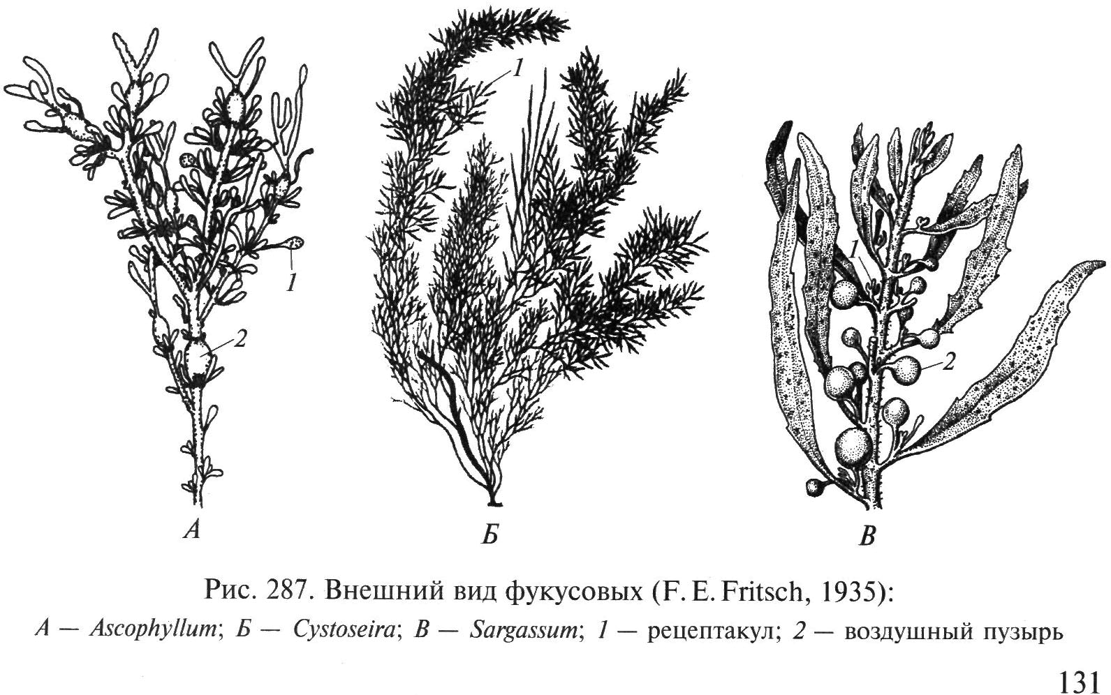
Rice. 34. Appearance of fucus algae: A – Ascophyllum; B – Cystoseira; IN – Sargassum; 1 – receptacle, 2 – swim bladder
a branched thallus with flat branches having a longitudinal rib, which is attached to the stones with a conical base. Fucus is used as fertilizer, as livestock feed, for the production of feed flour, alginates, and is used in medicine for the treatment of goiter, weight correction, etc.
Order Ectocarpaceae–Ectocarpales. The algae that are included here are microscopic, free-living, epiphytes, epizoites or endophytes. The reproductive organs are represented by single-locular (sporangia) and multilocular (gametangia) sporangia; they are long-cylindrical, short-cylindrical or pod-shaped.
Genus Ectocarpus. Thallus in the form of soft brown bushes up to 2 cm in height. The bushes are formed by single-row, alternate, dichotomous or irregularly branched threads. Multilocular sporangia are pod-shaped, formed on the side of the lateral branches on a 1–3-cell stalk. The species are distributed in seas of various latitudes, in clean and polluted waters, in the littoral zone and in the upper part of the sublittoral zone. Epiphytes are also found in the fouling of anthropogenic substrates.
Ecology
The vast majority of brown algae live in marine waters. IN fresh waters Only 8 species are found. There are both annual and perennial species, the age of which can reach 15–18 years. Laminaria algae Nereocystis And Macrocystis, which live along the Pacific coast of North America, have the maximum size for all algae - up to 30–40 m in length. They form giant underwater forests in the seas of the Far East. Brown algae attach to a wide variety of substrates - rocks, stones, gravel, shells and shells of sea animals, and other algae. Some small forms of brown algae live inside the tissues of other algae as endophytes. Most brown species live in an attached state. Thalluses torn from the ground are carried by the current to calm places with a muddy bottom and continue to exist there. Species with air bubbles on the thallus, detached from the ground, float to the surface, forming large floating aggregations, especially in areas with a stable circular current, as in the Sargasso Sea. They exist in such clusters quite for a long time and reproduce only vegetatively.
Brown algae are widespread in all seas, but they reach their greatest development in the seas of temperate and subpolar latitudes. Their large thalli grow attached to rocks and stones, and in calm places near the shore and at great depths they can even rest on the valves of mollusk shells and gravel. They can be found at different depths - from the littoral zone, where during low tide they are out of water for hours, to 40–200 m. In the area of the Hawaiian Islands, species of the genus were found at a depth of 180 m Sargassum, A Kelp V in the Adriatic Sea was found at a depth of 200 m. The most abundant thickets of brown algae are observed at a depth of 6–15 m, where there are the best lighting conditions and constant movement of water, which brings nutrients to their thalli and limits the settlement of herbivorous animals - phytophages.
Class Golden algae –Chrysophyceae
Unicellular, colonial or multicellular organisms, usually having a flagellar stage in their life cycle. Cells are mononuclear, with one or more chloroplasts. Stigma is usually present (Fig. 36). Flagella 1–2, unequal. The contractile vacuole is located at the front of the cell. The outer covers of the cells of these organisms are very diverse. In the most primitive species, the cell is covered with a delicate periplast, which allows the formation of protrusions of various shapes (rhizopodia, pseudopodia); in others, the plasmalemma is covered with a hard cellulose membrane, sometimes very slimy. The houses of golden algae come in different shapes: vase-shaped, spherical, ovoid, cylindrical; with one or more holes. The cage is attached to the base of the house with a flexible leg or can be free. In some chrysophytes, calcareous formations - coccoliths - of various shapes and sizes are deposited on the cell surface, and silicon flagellates have an internal silicon skeleton. The diversity of the structure of these skeletons makes it possible to use the remains of various forms of fossil silicoflagellates to determine the age of the geological rocks containing them. In more highly developed representatives of the golden algae department, the cells are covered with a shell consisting of silicon scales (sometimes bearing spines), or are enclosed in houses through the openings of which flagella or pseudopodia emerge.
The houses of golden algae species are vase-shaped, spherical, ovoid, cylindrical, with one or several holes. The cage is attached to the base of the house with a flexible leg or can be free.
Pigments - chlorophylls A And With, fucoxanthin. Chrysophytes are characterized by the highest fucoxanthin content among heterokonts (55-93% of all carotenoids).
Most species are freshwater phototrophs, but heterotrophs and phagotrophs are also found.
Reserve substances are chrysolamine and oils.
Among unicellular forms of golden algae, freshwater planktonic species predominate, and among multicellular forms, bottom or epiphytic species predominate. They are found mainly in cool sea and fresh water bodies. About 360 species are known.

Rice. 36. Golden algae (after: S. Hoek van den et al., 1995): A- cell structure; B– radicular system; IN– tripartite mastigonema; G– section of part of the chloroplast; 1 – basal body; 2 – basal swelling; 3 – short flagellum; 4 – stigma; 5 – rhizoplast; 6 – core; 7 – nuclear membrane; 8 – nucleolus; 9 – mitochondria; 10 – vacuoles; 11 – mucus; 12 – plasmalemma; 13 – vesicles; 14 – mucous body; 15 – lipids; 16 – chloroplast; 17 – Golgi apparatus; 18 – contractile vacuole; 19 – long flagellum: 20 – 1st root; 21 – 2nd spine; 22 – 3rd spine; 23 – 4th spine; 24 – chloroplast nucleoid; 25 – HES; 26 – chloroplast shell; 27 – encircling lamella; 28 – lamella; 29 – long lateral filament; 30 – short lateral filament; 31 – terminal filament; 32 – tubular part of the mastigonema
Thallus structure in golden algae it is most often monadic, but can be very diverse: coccoid, palmelloid, plasmoidal, amoeboid, filamentous, parenchymatous (Fig. 37).

Rice. 37. Golden algae (according to: L.L. Velikanov et al., 1981): A – Ochromonas: 1 – appearance, 2 – cyst; B – Chromulina: 1 – appearance, 2 – cyst, 3 – scheme of film formation from cysts; IN – Dinobryon: 1 – general view of the colony, 2 – cyst; G – Chrysameba; D – Gibberdia; E – Hydrurus: 1 – general view of the colony, 2 – tip of the branch, 3 – zoospore
Reproduction. Golden algae have three methods of reproduction: vegetative, asexual and sexual.
Vegetative propagation occurs by longitudinal cell division or disintegration of a colony into parts due to cell division in one, two or three directions.
Sexual process(cells of golden algae are diploid) – isogamy or conjugation. As a result of the sexual process, as well as under unfavorable conditions, cysts with a thick shell containing silicon are formed.
Asexual reproduction carried out by single- or biflagellate zoospores, which develop in ordinary vegetative cells or zoosporangia. Sexual process: holo-, iso- and autogamy. Hologamy is a type of sexual process in which two cells similar to vegetative ones merge at the anterior ends into a binuclear zygote, which then turns into a silicified cyst.
Taxonomy
From the Golden algae at the end of the last century, based on the results of molecular genetic studies, the class Sinuraceae was isolated. The class Pheotamniaceae is also distinguished - from Yellow-green and Golden algae. Therefore, the class Chrysophyceae has shrunk in size and currently contains about 360 species classified in three orders: Chromulinales, Hibberdiales and Hydrurales. The shape, structure and number of flagella, as well as the pigment composition and body structure are important systematic characters.
Order Chromulinaceae – Chromulinales. This order includes organisms with monadic, palmelloid and amoeboid types of thallus differentiation. Monad cells with one flagellum visible under a light microscope.
Genus Chrysameba(Fig. 37, A) are freshwater amoeboid algae; they have a flagellar stage in their life cycle. The zygote turns into a stomatocyst.
Genus Chromulina(Fig. 37, IN) - single-celled free-swimming organisms that live primarily in fresh waters. The cells are naked; there are one or two golden plastids.
Genus Dinobryon(Fig. 37 , G)– unicellular and colonial, free-swimming or attached representatives. Monads are located in vase-shaped houses formed by cellulose microfibrils. In addition to cellulose, the house also contains a large number of amino acids. The bush-like shape of the colonies is associated with the method of vegetative cell division, when one of the daughter cells, leaving the parent house, attaches to its opening. Rotating around its axis, it forms its own house. Both daughter cells can leave the parent house. The cells contain 1-2 chloroplasts; an ocellus and 2 contractile vacuoles are visible at the anterior end of the cell. Chrysolaminerin is located in a vacuole at the posterior end of the cell. Reproduction is vegetative and sexual.
Order Gibberdiaceae–Hibberdiales. Representatives of this order are characterized by a unique pigment composition. In addition to fucoxanthin, they have an additional light-harvesting carotenoid pigment - antheraxanthin.
Includes childbirth Gibberdia, Chromophyton, Styloceras, Chrysopixis, Platytheca and others.
Genus Gibberdia has two stages in the life cycle: colonial palmelloid non-motile and unicellular monadic motile. Under light microscopy, only one flagellum is visible at the monadic stage. Characteristically, in addition to fucoxanthin, there is another additional light-harvesting carotenoid pigment - antheraxanthin.
Order Hydrurus–Hydraulics. Representatives of this order have a thallus with palmelloid and pseudoparenchymatous types of structure. Characterized by the presence of unique zoospores of tetrahedral shape. There is no peephole. Mitosis is semi-closed.
Includes childbirth Hydrurus, Celloniella, Federmatium, Chrysonebula and others.
Genus Hydrurus(Fig. 37, E) is the most differentiated among palmelloid algae. Its thallus consists of large, up to 30 cm long, mucous colonies that look like brown branched cords, often emitting a very unpleasant odor. In the colonies, a main trunk and side branches can be distinguished. Cells immersed in common mucus contain one cup-shaped chromatophore and several contractile vacuoles. The cells are loosely located along the periphery of the colony and more densely in the middle. The thallus can only grow through division of apical cells. During asexual reproduction, zoospores are formed in the cells of the lateral branches of the colony. Forms spherical cysts. It is found in mountain streams and rivers with cold water, where it attaches to hard substrates.
Class Sinura algae –Sinurophyceae
syn- together and ura- tail. This group of organisms was isolated from golden algae in 1987. Combines monadic single and colonial organisms, sometimes with alternation of monadic and palmelloid stages in the life cycle. The surface of the pectin shells is covered with a shell of silicon scales. Mitochondria with tubular cristae are usually located in the cytoplasm around the chloroplast. There is one nucleus, usually two chloroplasts surrounded by four membranes. The lamellae are trithylakoid; there is a girdle lamella. The main pigments are chlorophylls A And With, β -carotene and fucoxanthin. There is no stigma. Cells usually have 2 unequal flagella. The long feathery flagellum is directed forward. The short, smooth flagellum, which is sometimes greatly reduced, is directed posteriorly.
Cells reproduce mainly by longitudinal division. Colonies, disintegrating, give rise to young colonies. In some species, the sexual process is described in the form of isogamy. Moreover, in contrast to golden algae, the fusion of isogametes in sinurian algae occurs not at the front, but at their rear ends. Endogenously, silicified cysts with pores are formed, similar to those of chrysophytes.
Mostly planktonic forms, palmelloid stages are part of the benthos. Most species of sinuric algae are phototrophic freshwater organisms.
Taxonomy.
The class Sinura algae includes 7 orders: Chloramoebales, Synurales, Rhizochloridales, Ochromonadales, Heterogloeales, Parmales and Thallochrysidales.
Order Sinuraceae – Synurales. Monadic forms, pectinous cell membranes are usually covered with a shell of silica scales, cemented by organic matter into a solid case. One or two chloroplasts.
Genus Sinura(Fig. 38) - freshwater monad forms with two unequal flagella and two wall chloroplasts. The nucleus is pyriform, located in the front of the cell. Behind the nucleus there is one large vacuole, and at the rear of the cell there are several small contractile vacuoles. The cells are covered with scales, like tiles. Reproduction is most often vegetative; for some, the sexual process is known. Forms colonies (Fig. 39, A). Cysts often appear in all cells of the colony. With mass development Sinura may give the water an unpleasant odor.

Rice. 38. Appearance of Sinura cells
Order Rhizochloridales – Rhizochloridales. Predominantly freshwater organisms with a rhizopodial type of thallus differentiation, living mainly in fresh water bodies.
Genus Mallomonas(Fig. 39, B-G) – a unicellular monad with one clearly visible flagellum and one bifurcated chloroplast.

Rice. 39. Appearance of sinur algae (according to: G.A. Belyakova et al., 2006): A – dividing colony Sinurs; B, C – formation of statospores and G – appearance Mallomonas
The cell is covered with imbricated and spiral scales, some of them have needle-like spines (Fig. 39, G). The presence of statospores is characteristic (Fig. 39, B, C). For a number of representatives, the sexual process is described - hologamy.
Genus Myxochloris lives in the cells of leaves of sphagnum mosses, has the appearance of a large multinucleate plasmodium. In the fall it forms cysts that germinate in the spring. Zoospores or amoebas emerge from them, penetrating the empty cells of the leaves and merging there into plasmodium.
Order Chloramoebales - Chloramoebales. Includes monadic representatives. Found in salt and fresh waters.
Genus Heterochloris – its cells can change shape, forming pseudopodia. This phenomenon is inherent in many representatives of the order, as well as the tendency towards an animal way of eating. The cell contains several chloroplasts, oil droplets and chrysolamine. In the front of the cell there are contractile vacuoles, in the center there is one nucleus. Reproduces vegetatively by longitudinal cell division.
Order Ochromonadales - Ochromonadales. Combines naked forms with two unequal flagella. Freshwater and marine forms.
Genus Ochromonas includes single-celled monads with two unequal flagella. The cells are clothed only with plasmalemma. At the anterior end of the cell there is a contractile vacuole and an ocellus, at the posterior end there is a vacuole with chrysolaminarin. It reproduces vegetatively; the cleavage furrow begins at the anterior end of the cell between two pairs of flagellar bases. Species of the genus are common in oligotrophic fresh waters, but there are also marine representatives.
Order Heterogloeales - Heterogloeales. Includes algae with palmelloid type of thallus differentiation. Representatives of the order are found more often in fresh than in salt waters.
Genus Helminthoglea lives in brackish waters. This colony, sitting on an expanded base, consists of branching mucous cords. In this mucus, protoplasts are randomly located, each of which is surrounded by its own mucous membrane.
Class Pheothamnia algae – Phaeothamniophyceae
The name of the class comes from the type genus Phaeothamnion(from Greek phaeos- dark bush). Representatives of this group of organisms were separated into a separate class of yellow-green and golden algae in 1998 based on an analysis of the sequence of ribosomal genes and features of cell ultrastructure. Pheotamnia algae are characterized by a unique combination of pigments: fucoxanthin with heteroxanthin, the absence of violaxanthin.
Features of the cell structure: the absence of vacuoles with chrysolaminarin and the absence of endogenous cysts with silica walls.
The class includes unicellular, colonial and multicellular organisms with coccoid, palmelloid and filamentous types of thallus structure. Flagellar stages with two unequal flagella. Flagella lateral or subapical. Stigma occurs in zoospores. Pigments - chlorophylls A And With, β -carotene, fucoxanthin, diadinoxanthin, diatoxanthin and heteroxanthin. The main reserve product is paramylon ( β -glucan). There is a cell wall, during cell division a daughter wall is formed inside the parent wall.
In pheotamniaceae, the main methods of reproduction are only vegetative and asexual. Asexual reproduction is carried out by autospores or zoospores. Sexual reproduction is unknown.
Species of Pheotamnia algae live exclusively in fresh waters. Taxonomy.
Currently, the monophyletic nature of the class of pheotamnia algae has been confirmed, and among ochrophytes it is closer to brown and yellow-green algae than to golden algae. Currently, about 30 species of these algae are known, classified as one order Pheothamniales - Phaeothamniales.
Genus Theotamnion represents attached branching threads up to 1 cm in height (Fig. 40). The cells are cylindrical, irregular, widened upward, along

Rice. 40. Appearance Theotamnion.
length two to three times the width. During cell division, the material of the parent wall is spent on the formation of a layered cover surrounding the cells of the filament. There are vesicles along the cell periphery under the plasmalemma. They resemble physodes - formations found in the cells of brown algae. The cells contain from one to several disc-shaped plastids of olive-brown color. Zoospores are formed 1–2, less often 4–8 in one cell. In zoospores, flagella are attached to the side. The position of the flagellar roots Theotamnion resembles that of yellow-green and brown algae. It also lacks a rhizoplast. During the life cycle, cysts can form, but there is no silicon in their walls. Cysts germinate as zoospores. Settles as an epiphyte on filamentous algae. Inhabits stagnant and slow-flowing fresh water bodies.
Class Raphid algae –Raphidophyceae
The name of the class comes from the Greek. rhaphid – needle. Unites unicellular biflagellate organisms lacking a cell wall. Mostly freshwater organisms with flattened cells (Fig. 41).
The cells have flagella of unequal length, the forward-directed flagellum is feathery and longer, the backward-directed flagellum is smooth and shorter. There is no transition spiral. Chloroplasts are small, numerous or, less commonly, 1-2 per cell, surrounded by four membranes, two of which are CES membranes. Thylakoids are collected in stacks of three. The core is large, surrounded by a ring of dictyos. Mitochondria with tubular cristae. There are contractile vacuoles. The cytoplasm is often vacuolated. The cells are naked, surrounded only by plasmalemma. There is usually no stigma (eye).
Photosynthetic pigments - chlorophylls A And With, β -carotene, voucheriaxanthin, dinoxanthin, diadinoxanthin, heteroxanthin.
Spare assimilation products are fats and oils, less often starch and chrysolamine.
They also live in sphagnum bogs and other habitats with acidic and neutral water reactions. Some species are found in brackish and marine waters.

Rice. 41. Appearance of raphid algae.
Taxonomy.
For a very long time, this group of algae, due to their green color, the presence of a pharynx and a number of other structural features, was considered as a class of Chloromonas among Euglenophytes. But the data of cytology, biochemistry, physiology and molecular biology showed their undoubted belonging to the Heterokontae (Ochrophyta) algae department and the monophyletic nature of the Raphid algae group.
About 25 species are known from the only order Huttonellaceae– Chattonellales.
Genus Heterosigma(Fig. 42, A) includes marine flagellates. Oval-shaped cells with flagella shifted to side, plastids are located along the periphery of the cell. There are no trichocysts. They swim, making rotating movements around the longitudinal axis of the body. May cause toxic algal blooms in coastal waters.

Rice. 42. Raphid algae (according to: R. E. Lee, 1999): A – Heterosigma; B – Goniostomum; 1 – chloroplast; 2 – mucocyst; 3 – contractile vacuole; 4 – trichocyst; 5 – core
Genus Goniostomum(Fig. 42, B) - mobile dorsoventrally compressed monads. The dorsal side is curved, the ventral side is flattened. A groove at the anterior end leads into a triangular pharynx, from which two long flagella emerge; their length is comparable to the length of the cell. Plastids are located along the periphery of the cell. Trichocysts are located under the plasmalemma. Lives in fresh waters with an acidic pH value.
Class Eustigma algae –Eustigmatophyceae
The name of the class comes from the Greek. eu- “good” and stigma- “mark”, “spot”. The class unites naked unicellular, less often colonial, organisms with a predominantly coccoid structure.
One or more cores. There is usually one chloroplast surrounded by 4 membranes. The flagellum is usually one anterior pinnate and a second basal body or, less commonly, two unequal flagella. The stigma (eye) is present, located outside the chloroplast, its granules without membranes.

Rice. 43. Appearance of eustigma algae
The chloroplast is usually single, large, multilobed, cup-shaped or parietal, yellow-green in color. The thylakoids are collected in lamellae of three; there is no girdle lamella.
Main pigments: chlorophyll a, β-carotene, violaxanthin and voucheriaxanthin. The pyrenoid is usually present only in vegetative forms.
Reserve substances are oils and substances of unknown nature that are deposited as solid material outside the plastids. No starch is formed.
They live in fresh waters, less often found in the seas and in the soil.
Taxonomy.
Previously, this group of organisms was classified as yellow-green algae. Currently, Eustigma algae are considered as a class in the Heterocontophytes department. About 35 species are known, mostly freshwater, attributed to the only order Eustigma - Eustigmatales.
Genus Evstigmatos(Fig. 43) – unicellular small spherical algae. The cell wall is smooth, solid, without ornament. The yellow-green chloroplast is single, lobed, parietal, with stalked polyhedral pyrenoids. Large central vacuole with red contents. It reproduces by 2 or 4 autospores or bottle-shaped zoospores with one incoming flagellum, a large stigma located outside the chloroplast at the anterior end, and a single posterior chloroplast without a pyrenoid. A common component of soil floras. Known from New Zealand, Austria, Iceland, and the Arizona basins as "mustard algae."
Class Yellow-green algae –Xanthophyceae
Yellow-green algae include algae whose chloroplasts are colored light or dark yellow, very rarely green and only sometimes blue. The color of thalli is determined by the presence of the following pigments in the chloroplasts of cells - chlorophylls A And With, β -carotene and xanthophylls. The predominance of the latter determines the unique color of yellow-green algae. In addition, paramylon, oil droplets, and only in some species, lumps of leukosine and volutin accumulate in cells as the main assimilation product. Yellow-green algae do not produce starch. A distinctive feature of yellow-greens is the presence of a monadic structure in vegetative cells and two unequal flagella in zoospores. The cell wall contains cellulose, glucose and uronic acids. The cell wall often consists of two parts.
Reproduction is vegetative, asexual and sexual.
Widely distributed in fresh waters. Rarely found in sea, brackish waters and soil.
Previously, the class Yellow-green algae was called Tribophycean algae after the type genus Tribonema (from the Greek. tribe – skillful, cunning and nema – a thread). About 450 species are known.
Yellow-greens are characterized by significant morphological diversity. Among the numerous representatives of this department, almost all the main types of body structure are found: amoeboid, monadic, palmelloid, coccoid, filamentous, heterofilamentous, lamellar and siphonal (Fig. 44 – 46). Thallus unicellular,

Rice. 44. Appearance of yellow-green algae: 1, 2 – Charatiopsis, 3 – Centritractus, 4 – Ophiocytium
colonial, multicellular and noncellular. The cell membrane is dense, pectin and cellulose, consisting of parts tightly overlapping each other or of two leaves. Silica or lime is deposited in the shell. Mostly immobile forms. Among unicellular species there are mobile forms that lack a dense shell and are equipped with flagella, lobopodia and rhizopodia.

Rice. 45. Appearance of xanthococcal yellow-green algae: 1–3 – Botridiopsis, 4 – Tetrahedriella, 5 – Pseudostaurastrum, 6 – Goniochloris, 7, 8 – Bumilleriopsis
Most yellow-green – immobile organisms. In motile individuals, movement can be carried out using flagella or rhizopodia. Cells of various shapes: spherical, spindle-shaped, ellipsoidal, cylindrical, tetrahedral, sickle-shaped, pear-shaped, ovoid. Thalluses ranging in size from 0.5 – 1.5 µm ( Chloridella) up to several millimeters in diameter ( Botridiopsis) (Fig. 45, 1 – 3) and up to tens of centimeters in length ( Vaucheria) (Fig. 46, 3).

Rice. 46. Appearance of yellow-green algae: 1 – Tribonema, 2 – Heteropedia, 3 – Vaucheria, part of a filament with oogonium and antheridium
Most yellow-green species are phototrophs, but holozoic feeding by ingesting bacteria and small algae is also found. Yellow green algae are widespread in fresh waters. They are also common in soil, less common in marine and brackish water bodies. The class includes aerobiont, planktonic, benthic and periphytonic forms. Epiphytes, epizoites, as well as intracellular symbionts in protozoan cells.
Regardless of the external structure, the internal structure of the cell of yellow-green algae is the same. In the protoplast, several yellow-green chloroplasts are usually observed, having a disc-shaped, trough-shaped, lamellar, less often ribbon-shaped, stellate or cup-shaped with solid or lobed edges. The color is due to the lack of fucoxanthin, which is responsible for the golden and brown color in other ochrophytes. Among other pigments they have β -carotene, voucheriaxanthin, diatoxanthin, diadinoxanthin, heteroxanthin. In motile forms, a red eye, or stigma, is usually located at the anterior end of the chloroplast. Few species have semi-submerged pyrenoids. The nucleus in a cell is usually one, small in size, but there are species with multinucleated cells. In some species, there are one or two contractile (pulsating) vacuoles at the front of the cell.
Monad representatives and motile stages (zoospores and gametes) have two unequal flagella. The exception is synzoospores Vaucheria, in which numerous pairs of smooth flagella of slightly different lengths are located on the surface. The short flagellum ends with an acroneme. Flagella are attached subapically to the cell. In the sperm Vaucheria attachment is lateral.
Species with amoeboid, monadic and palmelloid organization lack a cell wall, they are covered only by a cytoplasmic membrane and can easily change shape. Sometimes “naked” cells are located inside houses, the walls of which can be painted brown with manganese and iron salts. The vast majority of forms have a cell wall consisting of two parts. The cell wall is dominated by cellulose and also contains polysaccharides, consisting mainly of glucose and uronic acids. Young cells have a thin membrane, but with age it thickens. Iron salts can be deposited in it, the compounds of which color it in various shades of brown and red. Most often, silica is present in the cell wall, giving it hardness and shine. It can also be encrusted with lime and be sculptured in various ways (spines, cells, warts, bristles, denticles, etc.) Attached forms can form an outgrowth of the shell – leg with attaching sole.
In filamentous forms of yellow-green algae with bivalve shells, when the filaments disintegrate, the cell membranes fall apart into H-shaped fragments. These fragments are tightly connected halves of the membranes of two neighboring cells (Fig. 47). As filaments grow, an H-shaped fragment of the cell wall of two adjacent daughter cells is inserted between the two halves of the mother cell wall. As a result, each of the daughter cells is half covered with the old membrane of the mother cell and half – newly formed membrane.

Rice. 47. Scheme of the formation of a transverse partition between two daughter cells in filamentous yellow-green algae (according to: A.A. Masyuk, 1993): A– fragment of thread; B– laying of the girdle ring and formation of a transverse septum between two cells; IN– layering of bicuspid cell membranes; G– disintegration of the shell into H-shaped sections
Contractile vacuoles are present in motile representatives. Usually there are 1-2 of them per cell. The Golgi apparatus has a unique structure. Dictyosomes are small, containing 3-7 cisternae. There is one core, less often there are many of them; In coenotic species, the cells are always multinucleated.
Reproduction. Most species of yellow-green algae are characterized by vegetative and asexual reproduction.
Vegetative propagation carried out in various ways: dividing cells in half, disintegrating colonies and multicellular thalli into parts. U Vaucheria Special brood buds are formed.
At asexual reproduction A variety of spores can be formed: amoeboids, zoospores, synzoospores, autospores, hemizoospores, hemiautospores, aplanospores. Zoospores are “naked” and usually pear-shaped.
Sexual process– isogamy, heterogamy and oogamy – described in a few representatives. U Tribonemes gametes are similar in size, but differ in behavior - this is isogamy. U Vaucheria Oogamy is observed: receptacles for female gametes are formed on the threads – oogonia and male – antheridia.
IN unfavorable conditions formation of cysts is observed. Cysts (statospores) are endogenous, mononuclear, less often multinucleate. Their wall often contains silica and consists of two unequal or, less commonly, equal parts.
Taxonomy.
At the end of the XIX – beginning of the 20th century various genera of yellow-green algae were classified as green algae, which was primarily due to the color and morphological similarity of the thalli. Currently, the yellow-greens are considered as a class within the ochrophyte division.
About 450 are known modern species class Yellow-green algae, which are grouped into four orders: Botridiaceae, Michococcaceae, Tribonemaceae and Vaucheriaceae. The identification of orders is based on the type of differentiation of the thallus and the characteristics of the life cycle.
Order Botridiaceae – Botrydiales. The order includes species with a siphonal type of differentiation of the thallus, in which there is no oogamous sexual process.
Genus Botridium lives on the soil and has the appearance of green bubbles several millimeters in size, attached with the help of colorless rhizoids. The thallus is siphonal, contains numerous nuclei and plastids. The shell is multi-layered and lime can be deposited on it. Reproduction is asexual with the help of biflagellate zoospores, and the entire contents of the bladder disintegrate into mononuclear fragments. When there is a lack of moisture, it reproduces using aplanospores or forms thick-walled cysts. In some cases, the entire contents of the bladder are used to form one large cyst. In other cases, cysts form in rhizoids, where the contents of the bladder first move. Cysts germinate either directly into a new thallus or form zoospores. The sexual process is iso- and heterogamy. The zygote germinates immediately, without a dormant period. Common and widespread species in terrestrial habitats, found along the banks of streams, ponds or on soils devoid of vegetation.
Order Mischococcaceae–Mishococcales. Unicellular or colonial representatives with a coccoid type of differentiation of the thallus.
Genus Charatiopsis includes unicellular attached forms. During reproduction, it forms zoospores, aplanospores and thick-walled cysts (Fig. 44, 1-2).
Genus Ophiocytium(Fig. 44, 4) has elongated cylindrical cells, which can be straight, bent or spirally twisted, and may bear a spike at the end. The cell wall consists of two unequal parts, most of which are involved in cell growth, the smaller part is permanent and has the shape of a lid. Unicellular and colonial species, free-living or attached to the substrate with a small stalk. They reproduce by zoospores and aplanospores, and cysts are also found. They live in fresh waters.
Genus Mischococcus forms tree-like attached colonies. Branching is dichotomous and tetrachotomous. The cells are located in groups of 2 or 4 at the tops of the mucous branches of the colony. The cells are spherical to oval, with a thin or thick cell wall. Sometimes the cell wall is shiny and brown due to its impregnation with iron salts. Young single-celled organisms with a slimy disc-shaped base that serves as a fulcrum for attachment. After the spores are released, the protoplast of the mother cell turns into jelly and stretches, the length becomes 6 times greater than the width, and thus a cylindrical leg appears. The empty cell wall of the mother cell always becomes the base of the stalk. Asexual reproduction by zoospores and autospores. Autospores are attached to the upper edge of the mucous stalk. Subsequent cell divisions repeat the process and produce a tree-like colony. Sexual process – isogamy. They live in small fresh water bodies as epiphytes of filamentous algae. Known in central Europe and Asia.
Order Tribonemales – Tribonematales. Representatives have filamentous, heterofilamentous, pseudotissue and tissue types of thallus differentiation. The cell walls are either with H-shaped overlapping parts or solid.
Genus Tribonema– non-branching threads (Fig. 46, 1). The cells are cylindrical or barrel-shaped. The cell wall consists of two halves, which end up facing each other in the middle of the cell. The shells are often layered. Fragments of threads always end in empty halves of H-shaped fragments of the shell, shaped like a fork. The cells contain several yellowish-green plastids and no pyrenoids. Reproduction is vegetative (by fragmentation of filaments), asexual (zoospores and aplanospores) and sexual (isogamy), with aplanospores being formed more often than zoospores. Can form akinetes. They live in fresh waters, where they develop especially abundantly in the cold season.
Order Vaucheriales. All representatives have a siphonal thallus, oogamous sexual process and synzoospores.
Genus Vaucheria(Fig. 46, 3) has a thallus of noncellular structure; its thallus reaches a length of several centimeters and is attached to the substrate with the help of a colorless rhizoid. There are no partitions in the filaments, most of the thallus is occupied by a vacuole, and numerous nuclei and plastids are located along the periphery in the cytoplasm. Filaments with apical growth and sparse lateral branching. The septa are formed when the thallus is damaged and to separate the reproductive organs. Asexual reproduction is carried out by aplanospores, synzoospores, and akinetes. Synzoospores are formed one at a time in the zoosporangium, which is separated from the vegetative cells by a septum at the end of the filament. Zoospores are multinucleate and multiflagellate. The sexual process is oogamy. The zygote becomes covered with a thick shell and, after a period of rest, grows into a new thallus.
Kinds Vaucheria widely distributed in fresh, brackish and marine waters, as well as in terrestrial habitats. They are found on all continents, including Antarctica. They form grassy-green or dark green tangled masses - so-called mats, smooth, creeping or cushion-shaped. Aquatic, semi-aquatic, terrestrial forms. They live in a variety of habitats: seas, estuaries, estuaries, salt marshes, mangroves, streams, canals, lakes, ponds, arable lands and swamps.
Meaningheterokont algae
From the Heterokont algae department, brown algae are of greatest importance for natural ecosystems and for humans.
Brown algae - main source of organic matter in the coastal zone of the seas. Their biomass in the seas of temperate and subpolar zones can reach several tens of kilograms per square meter. Thickets of brown algae create conditions for the feeding and reproduction of many coastal animals and other algae. Charles Darwin, who observed kelp forests off the coast of South America Macrocystis, wrote: “I can only compare these huge underwater forests of the Southern Hemisphere with the terrestrial forests of tropical regions. And yet, if a forest were destroyed in any country, I don’t think that at least approximately the same number of animal species would die as with the destruction of this algae.”
Thickets of brown algae serve food place,shelter and breeding many animals. Figuratively speaking, brown algae provide other aquatic organisms with a “table, shelter and nursery.”
Brown algae is also widely used by humans. They are rich iodine and other microelements. The peoples of Southeast Asia traditionally use them for food, especially representatives of the order Laminariaceae, from which many different dishes are prepared. Feed meal, prepared from brown algae, increases livestock productivity; at the same time, the iodine content in eggs and milk increases.
From brown algae getalginates– salts of alginic acid. Alginates are widely used in various industries. These are non-toxic compounds with colloidal properties, so they are widely used in the food and pharmaceutical industries. Alginic acid and its salts are capable of 200–300-fold absorption of water, forming gels that are characterized by high acid resistance. In the food industry they are used primarily as emulsifiers, stabilizers, gelling and moisture-retaining components. For example, dry powder sodium alginate is used in the production of powdered and briquetted soluble products (coffee, tea, milk powder, jelly, etc.) for their rapid dissolution. Aqueous solutions alginates are used for freezing meat and fish products. In the world, up to 30% of the total volume of alginates produced goes to the food industry.
In the textile and pulp and paper industries, alginates are used to thicken paints and enhance the strength of their bond to the base. Impregnation of fabrics with some alginates gives them protective properties: waterproofness, acid resistance and increases mechanical strength. A number of salts of alginic acids are used to produce artificial silk. During World War II, a large amount of camouflage fabric and nets for residential and industrial buildings was produced from alginic acid and its salts in the USA and England.
Alginates are used in metallurgy: in foundries they improve the quality of molding earth. Alginic acid salts are used in the production of electrodes for electric welding, which make it possible to obtain higher-quality welds. Alginates are also used in the production of plastics, synthetic fibers, paints and weather-resistant building materials. They are used in the manufacture of high-quality lubricants for machines. In radio electronics, alginates act as a binding agent in the production of high-quality ferrites.
The most widely used is water-soluble sodium alginate, which is capable of forming viscous solutions. It is widely used to stabilize a variety of solutions and suspensions. Adding a small amount of sodium alginate to food products - canned food, ice cream - improves their quality. It is also used to make decorative cosmetics, creams and masks in the perfume industry.
In the pharmaceutical industry, alginates are used to coat tablets, pills, as component bases for various ointments and pastes, as gel carriers for drugs, in the production of soluble surgical sutures. In medicine, calcium alginate is used as a hemostatic agent and as a sorbent that removes radionuclides (for example, strontium). The annual production of alginates in the world exceeds 20 thousand tons.
Another important substance obtained from brown algae is the hexahydric alcohol mannitol. Mannitol is used as a sugar substitute for diabetics. In addition, it can be used as a plasma substitute for blood conservation. It is used to make tablets in the pharmaceutical industry. Mannitol is also used in production of synthetic resins, paints, paper, explosives, and leather tanning.
Fucoidans, obtained from brown algae are effective anticoagulants, even more active than heparin. Their use for the production of antitumor drugs and antiviral compounds is considered promising. Indeed, even at the lowest concentrations, fucoidans can inhibit the attachment of viruses to the cell surface. Fucoidans also have the ability to form extremely strong and viscous mucilages, which are used in the preparation of stable emulsions and suspensions.
The energy crisis, which has engulfed many countries around the world in recent years, has led to the need to search for new non-traditional energy sources. Thus, in the USA, the possibility of breeding giant kelp algae is being studied for this purpose. Macrocystis with subsequent processing into methane. It is estimated that from an area of 400 km 2 occupied by this algae, 620 million m 3 of methane can be obtained.
Heterokont algae from the classes Golden, Yellow-green, Sinuraceae, Raphidophyta and Eustigma algae, represented mainly by microscopic organisms, are widespread in fresh water bodies of all climatic zones globe, but are more common in temperate latitudes. Among golden algae, there are species that live in seas and salt lakes, and very few live in polluted waters. Golden algae reach their maximum development in the cold season: they dominate plankton in early spring, late autumn and winter. At this time, they play a significant role as producers of primary production and serve as food for zooplankton organisms.
Some golden algae, e.g. Uroglen And Dinobryon, developing in mass quantities, they can cause algal blooms. They produce aldehydes and ketones, which can give water an unpleasant odor and taste, a Uroglen– fatty acids toxic to fish.
Raphid algae are widely represented in the plankton of fresh water bodies, mainly with an acidic pH, especially in sphagnum bogs, and less often in large lakes. In fresh water bodies, local “blooms” can form Goniostomum. Raphid algae are also found in desalinated sea bays and puddles on the seashore, as well as in the open sea. When they develop en masse in coastal sea waters, they cause toxic “blooming” of water. Thus, off the coast of Canada during a “bloom”, the concentration of cells of the raphid algae Heterosigma can reach 30 million per 1 liter. Outbreaks of raphid algae often lead to the development of “red tides,” which are associated with fish kills. The cause of such “red tides” may be the types of births Hattonella, Olistodiscus, Heterosigma and Fibrocapsa.
Sinur algae, when developed en masse in fresh water bodies, can give the water an unpleasant odor ( Sinura). Pheotamnia algae are found in standing and slowly flowing fresh water bodies, where they settle epiphytically on filamentous algae.
Eustigma algae are found only in fresh water bodies or in soil.
Yellow-green algae are common on all continents, living primarily in fresh water and soil, as well as in terrestrial, brackish and marine habitats. Yellow-green algae inhabit clean and polluted waters, with different pH values: they can live in both acidic and alkaline waters. They are found mainly in clean freshwater reservoirs, less often in seas and brackish waters, preferring moderate temperatures, most often developing in spring and autumn, although there are species found throughout all periods of the year, including winter. Most often they can be found in accumulations of filamentous plants and among thickets of higher aquatic plants in the coastal zone of rivers, ponds, lakes and reservoirs.
The vast majority of yellow-greens are free-living forms, but intracellular symbionts – zooxanthels – are also found in protozoan cells. Marine chloroplasts form an interesting intracellular symbiosis Vaucheria with clam Elision. For nine months, this mollusk is capable of photoautotrophic carbon dioxide fixation in culture. This is the longest symbiosis of this type, when the symbiotic plastid is in direct contact with the cytoplasm of the animal. In nature, mollusk larvae feed on threads Vaucheria. As a result of phagocytosis, algae chloroplasts enter the cytoplasm of the mollusk epithelial cells. During this process, the chloroplast membrane becomes three-layered, and one outer membrane of the chloroplast endoplasmic reticulum (chloroplast endoplasmic reticulum) is lost. This phenomenon provides good evidence that during evolution, as a result of secondary symbiogenesis due to the loss of membranes, chloroplasts with three membranes could arise.
Yellow-green, golden and other heterokont algae are producers of oxygen and organic substances; they are part of food chains. Heterocontophytes participate in the self-purification of polluted waters and soils, the formation of sapropel, and in the process of accumulation of organic substances in the soil, affecting its fertility. They are used as indicator organisms in determining the status of water pollution; yellow-green algae are part of a complex of microorganisms used for wastewater treatment.
Control questions
Name the characteristic structural features of brown algae.
Features of the structure of brown algae thalli.
How do brown algae reproduce? What are monospores, tetraspores and zoospores, isogamy, heterogamy and oogamy?
What are the life cycles of brown algae? Reproduction of fucus and kelp algae.
Name characteristics and typical representatives of the orders of brown algae.
In what habitats are brown algae found? What is their significance in nature?
Economic importance of brown algae.
Name the characteristic structural features and typical representatives of golden algae.
What pigments and nutritional types are known in golden algae?
Reproduction and ecology of golden algae.
Name the characteristic features and typical representatives of yellow-green algae.
What pigments and nutritional types are known in yellow-green algae?
How do yellow-greens reproduce? Types of sexual reproduction: isogamy, heterogamy and oogamy?
Name the characteristic structural features and typical representatives of sinuric algae.
Reproduction and ecology of sinur algae.
Name the characteristic structural features and typical representatives of pheotamnia algae.
Reproduction and ecology of pheotamnia algae.
Name common features structure and typical representatives of raphidophyte algae.
Reproduction and ecology of raphidophyte algae.
Name the general structural features and typical representatives of eustigma algae.
Reproduction and ecology of eustigma algae.
The importance of heterokont algae in natural ecosystems.
All heterokonts have a similar structure of the flagellar apparatus. There are 2 flagella, and one of them has very characteristic tubular three-membered feathery outgrowths, or hairs - mastigonemes. It was the presence of two unequal flagella that served as the basis for naming this large group of aquatic organisms heteroflagellatesalgae, or heterokonts. In addition to differences in length, the flagella also differ morphologically: the main flagellum consists of an axis and ciliated hairs pinnately located on it, the lateral flagellum is smooth, whip-shaped. The composition of pigments – chlorophylls – is also similar A And With and numerous carotenoids, as well as the structure of chloroplasts. Chloroplasts are surrounded by four membranes, with the outer two membranes connected to the cell's endoplasmic reticulum.
As part of the department, Heterocontaceae (Hydroflagellates) algae unite several groups of algae in the rank of classes: Brown, Golden, Yellow-green, Raphidophyta, Sinuraceae, Pheotamniaceae, Eustigmaceae, some were previously considered as independent departments.
Class Brown algae– Phaeophyceae (Fucophyceae)
The name of the class “brown algae” reflects the color of their thallus (from the Greek. Rhaios– dark-skinned). The color is due to the presence of a large number of brown and yellow pigments - in addition to chlorophylls A And With And β -carotene in their chloroplasts there is an excess of xanthophylls, especially fucoxanthin.
Multicellular, predominantly large seaweeds, especially widespread in the cold waters of both hemispheres. Neither unicellular nor colonial forms are known among brown algae. About 1.8 thousand species are known.
Cell structure. The cell of brown algae species is eukaryotic: it has formed organelles with their own membranes: nucleus, mitochondria, plastids, etc. Green pigments are chlorophylls A And With– are masked by additional: orange carotenes and yellow-brown xanthophylls; Of the latter, fucoxanthin predominates. Brown algae cells contain from one to many plastids. Brown algae cells typically produce a tough cell wall on top of their outer cell membrane.
Most often there is one core, but sometimes there are several cores. In some representatives, as the age of the cell increases, the number of nuclei increases. There is always one nucleolus in the nucleus.
Chloroplasts of vegetative cells of brown algae have an eye (stigma) and pyrenoids, of which there are one or several; some species lack pyrenoids. The ocellus, consisting of 30–40 lipid globules, acts as a lens that focuses light onto the flagellar swelling, which is a photoreceptor. Zoospores and gametes of brown algae usually do not have an eye. Reserve substances are deposited in pyrenoids. Chloroplasts are most often small, disc-shaped, parietal. Their shape can be star-shaped, ribbon-shaped or lamellar. The shape of chloroplasts in brown algae cells can change as the cell ages. The chloroplast envelope consists of four membranes, with the two outer membranes connected to the endoplasmic reticulum of the cell.
The cellulose that makes up the cell wall of brown algae, sometimes called algulose, differs in properties from the cellulose of higher plants. Outside the cellulose wall there is a highly slimy pectin layer, formed mainly by alginic acid and its salts and compounds with protein substances. Alginic acid also found only in brown algae.
General characteristics.
All brown algae are phototrophs. Products of photosynthesis - specific carbohydrates chrysolaminerin,alcohol mannitol And lipids. Lipids accumulate in the form of oil droplets. Chrysolaminerin is deposited outside the chloroplast: it is located in special vacuoles in the cytoplasm near the pyrenoid. Mannitol performs a reserve as well as an osmoregulatory function; its concentration inside the cell depends on changes in the salinity of the water.
Young cells of brown algae contain small and numerous vacuoles, which become larger with age due to fusion. In the cytoplasm there are special formations - physodes, which contain phlorotannins. These substances are known only from species of brown algae. The function of phlorotannins may be related to the protection of algal thalli from being eaten by animals, for example, mollusks. Phlorotannins from brown algae probably inhibit the settlement of epiphytic algae and animals on the surface of the thallus. It is assumed that phlorotannins are involved in protection against radiation damage and in the selective absorption of heavy metal ions from the aquatic environment. Colorless phlorotannins oxidize in air to form the pigment phycaffeine, which gives dried brown seaweed its characteristic dark color.
The structure of the thallus. Among the species of brown algae there are no unicellular or colonial forms; all individuals are multicellular. The shape of brown algae thalli is the most diverse. These are threads, creeping or vertical, single-row or multi-row; massive solid or imbricated crusts and crusts; thin mucous plates or dense leathery plates on the stem, simple or dissected; bubbles and bags; tubes and cords; variously branched thin bushes or powerful bushes. Attachment of thalli is carried out using rhizoids or soles. To maintain an upright position, large benthic forms of brown algae form air (swimming) bladders filled with gas (Fig. 29).
Rice. 29. Appearance of brown algae: 1 – Fucus; 2 – Saccharin; 3 – Alaria; 4 – Sargassum
Among the species of brown algae, the thalli of representatives of the Laminaria and Fucus orders are most complex. They have signs of tissue differentiation with specialization of thallus cells (Fig. 30). In their thalli one can distinguish bark, consisting of intensely colored cells and core, consisting of colorless cells that are often collected in threads. In Laminariaceae, sieve tubes and tubular threads are also formed in the core. The core performs not only a transport function, but also a mechanical one, since it contains threads with thick longitudinal walls. Between the bark and the core of many brown algae there may be an intermediate layer of large colorless cells.
Height thallus in representatives of brown algae is most often intercalary (intercalary) and apical (apical), less often basal. Intercalary growth may be diffuse or there may be a growth zone. In large forms, the intercalary meristem, or an actively dividing group of cells, is located at the transition point of the “petiole” to the “leaf” blade. Large algae also have a meristematic zone on the surface of the thallus, the so-called meristoderm (a kind of analogue of the cambium of higher plants).
Reproduction in brown algae, vegetative, asexual and sexual. Gametes are formed in multilocular gametangia, zoospores - in 
Rice. 30. Internal structure Laminaria(according to: L.L. Velikanov et al., 1991): A– longitudinal section of the stem; B– cross section of the stem; IN– section of a plate with zoosporangium sori
unilocular sporangia.
Vegetative propagation in a number of species of brown algae can be carried out by sections of the thallus; in species sort of Sphacelaria– brood branches (brood buds); in species of the genus Fucus- a group of cells on the sole that are capable of differentiation into a new thallus.
Asexual reproduction of brown algae usually occurs using zoospores. Some species reproduce by immobile tetraspores or monospores. Spores of asexual reproduction are formed as a result of meiosis and subsequent mitoses in unilocular sporangia. The sexual process is isogamy, heterogamy and oogamy. Gametes are formed in polygonbottom gametangia- special groups of cells. One gamete is formed in each “nest” of gametangium.
Known for brown algae sex pheromones– soluble substances that coordinate the activity of cells during reproduction. Pheromones, of which at least ten are known, can either stimulate the opening of antheridia or attract male gametes to female ones. They are also involved in species isolation. It is believed that pheromones are formed in most species of brown algae that have a sexual process.
Life cycle. Brown algae have two main types of life cycles. One type is haplodiplobiont. Sporophytes are diploid, i.e. all cells contain a complete set of chromosomes. Spores of asexual reproduction are formed in single-locular sporangia. When spores form, reduction division occurs—meiosis. This reduction is called sporical. The cells (spores) formed after meiosis are haploid, i.e. contain half the set of chromosomes. Haploid zoospores and tetraspores germinate into a haploid gametophyte, on which gametes are formed in multilocular gametangia. The diploid zygote formed after the fusion of gametes grows into a diploid sporophyte.
The other type is diplobiont; sporophytes, spores and gametophytes are diploid; reduction division occurs during the formation of gametes. This reduction is called gametic. After the fusion of haploid gametes, a diploid zygote is formed and grows into a diploid sporophyte. This type is common in kelp species (Fig. 31).

Rice. 31. Life cycle Sugars(according to: L.L. Velikanov et al., 1991): 1 – sporophyte, 2 – sori of zoosporangia, 3 – zoospores , 4 – gametophyte, 5 – antheridium, 6 – oogonia, 7 – antherozoid, 8 – zygote (fertilized egg)
On a macroscopic sporophyte Sugars, in the upper part of the plate, developing in stripes sori(groups of) zoosporangia. In zoosporangia, after reduction division, zoospores with two unequal flagella are formed. They grow into microscopic gametophytes: male with antheridia and female with oogonia. An egg fertilized by antherozoids immediately, without a dormant period, germinates and forms a sporophyte. This is the life cycle Sugars ends.
Fucus algae do not have sporophies. U Fucus male and female genital organs - antheridia with spermatozoa and conceptacles with oogonia and eggs - are located in the recesses of the thallus - scaphidia, male and female skafidia can be on the same recipecule or in different recipes. Oogonia sit on a stalked cell , arising on the wall conceptacle(Fig. 32, B, D). The oogonia cell undergoes three divisions

Rice. 32. Reproduction Fucus(according to: B.Fott, 1971; W.Braune et al., 1974): A – fragment of thallus (1 – air bubble, 2 - prescription); B– incision through the receptacle; IN– section of female scafidium; G– oogonia; D– release of eggs; E– section of male scafidium; AND– twig with antheridia; 3 – sperm output
nuclei, the first of which is reduction, resulting in the formation of 8 eggs in the oogonia.
Antheridia are usually formed on branched filaments that support them - paraphyses(Fig. 32, E,AND). As a result of several divisions, 64 sperm are formed. Sperm Fucus have an eye located inside a reduced chloroplast. At the anterior end of the sperm cell, 13 microtubules form hobocurrent- a special structure that is believed to be involved in the determination of the female sexual attractant.
Sperm are attracted to the egg Fucus nonspecific pheromone fucoserratene secreted by the egg. Recognition at the species level is associated with specific oligosaccharides on the egg and sperm. On the surface of the egg there are various areas, each of which contains special organic compounds - glycoproteins. Spermatozoa contain glycoproteins organized into regions on the membrane and processes of the anterior flagellum, as well as on the surface of the sperm itself. When meeting an egg, the sperm moves over the shell of the egg, as if examining it with the help of its anterior flagellum. If the glycoproteins of the sperm are complementary to the glycoproteins of the egg, only then does recognition of the sperm by the egg occur.
Taxonomy
Fossil finds that may be related to brown algae are known from the Middle Silurian (about 425 million years). But these finds also have similarities with some modern green and red algae. Fossil finds that can definitely be associated with modern brown algae date back to the Miocene (5–25 million years). These algae reminiscent of modern Dictyota. The age of brown algae, determined by molecular methods, is 155–200 million years.
The type of organization of the thallus, the presence or absence of a pyrenoid, the method of growth, the type of sexual reproduction (isogamy, heterogamy, oogamy) and life cycle are used to distinguish the orders of brown algae. In recent years, the brown algae system has been actively reconsidered. In different classification schemes, the Class Brown algae includes from 7 to 14 orders.
Order Desmarestiaceae–Desmarestiales. The algae are bushy, multi-branched, with a ridged, flat or flattened thallus. Attached to the substrate with a well-developed sole. Hairs develop on the surface of the thallus.
Genus Desmarestia (Fig. 33). Brown or olive bushes with a well-defined main shoot, up to 0.8–1 m in height. Attached with a cone-shaped sole. Branching in 3–4 orders, alternate or opposite. The branches of the last orders are flat or, less commonly, thread-like. The species are marine, live in arctic and temperate latitudes at a depth of 1–20 m, form thickets or grow singly.

Rice. 33. Desmarestia (b. Desmarestia)
Order Ralphsiaceae–Ralfsiales. Representatives of the order have a thallus in the form of crusts of various structures or consist of a basal crust and vertical shoots.
Genus Analipus. The thallus consists of a perennial crust-like basal part and annual, olive-brown, vertical shoots extending from it, up to 40 cm high. Multilocular sporangia are cylindrical in shape and two-row in structure. Unilocular sporangia are oval and develop among assimilative branches. Species of the genus are widespread in the seas of temperate latitudes, forming belts in the tidal zone.
Order Dictyotaceae–Dictyotales.Species of the order are characterized by apical growth. Vegetative cells contain numerous disc-shaped chloroplasts without pyrenoids. The thallus is parenchymatous, characterized by an isomorphic change of generations. The sexual process is oogamous, asexual reproduction is by immobile spores, spermatozoa have only one flagellum. The flagellum is long, with feathery projections and directed forward. Representatives of the order are found mainly in tropical and, less often, in temperate seas.
Genus Dictyota (application, 4A). It has a thallus in the form of flat olive-brown dichotomously branched bushes up to 11–15 cm high. The branches are linear, narrow, have no veins, with forked tips. Tetrasporangia form sori on the surface of the thallus, which have an indefinite or ring-shaped shape. Distributed in sea waters of temperate and warm latitudes.
Order Laminariaceae–Laminariales. Laminariaceae have a thallus consisting of a plate, a stem and attachment organs - the sole or rhizoids. The plate is smooth or wrinkled, whole or dissected, with or without holes, folds, boules, length from several tens of centimeters to 20–40 m. The stem is branched or more often unbranched. Sporophytes are annual or perennial, some species are 11–18 years old. In the bark of many kelp there are mucous canals.
The most popular representatives of brown algae are kelp (species of the genera Kelp or Saccharine), or “seaweed,” which are found primarily in the Pacific Ocean. They are found from the surface (edge) of the water to a depth of 200 m. Laminaria grow preferably at a depth of 2–20 (30) m, in places with constant movement of water, attaching to stones and rocks. Laminaria form algal belts along the coast of the seas of the Far East. The most powerful thickets are found near the Kuril Islands and about. Hokkaido, where about half of the world's kelp resources are concentrated. The annual global harvest of kelp algae reaches 2 million tons of wet weight, with more than 1 million tons coming from mariculture production in China.
Genus Saccharina (kelp). The thallus is from 0.5 to 5–10 m long, has a thick shiny plate, an elastic stem and densely intertwined rhizoids, with which it is attached to various solid substrates. The edges of the plate are smooth or wavy, and there are often bulges and folds on the surface of the plate. Clusters of sporangia - sori - are formed on both sides of the plate; after the spores are released, the upper part of the plate is destroyed. Lives from two to several years. Saccharins are valuable industrial raw materials and objects of mariculture. Japanese saccharine forms thickets in the northern half of the Sea of Japan, as well as off the southern and southeastern coast of Sakhalin and the South Kuril Islands. Japanese saccharine is the most valuable commercial species, it has excellent taste and is used for preparing a variety of dishes, canned food, and confectionery.
Order Sphacelariaceae–Sphacelariales.
Brown algae belonging to this order have a branching thallus with apical growth. Branches with hairs. Vegetative cells have numerous chloroplasts without pyrenoids. The life cycle of the majority is with an isomorphic change of generations, the sexual process is isogamy or heterogamy. Algae form small brown tufts up to 3 cm in height and branched bushes up to 20 cm in height on stones or other algae; there are endophytic representatives.
Genus Sphacelaria. It has thin filamentous thallus. Branching threads form small, up to 0.5–3 cm in height, brown tufts. Vegetative propagation is carried out by specialized, often three-rayed brood branches - propagules. The sexual process is isogamous. Female gametes secrete the pheromones ectocarpen and multifiden. Species of the genus are distributed in the seas of both polar and tropical latitudes.
Order Scytosiphonaceae–Scytosiphonales. Representatives with an intestinal, bladder-shaped, lamellar or saccular, often hollow thallus up to 25 cm in length. The bark is single-row or double-row, the inner part (wall) of the thallus is formed by several layers of large cells. On the surface of the thallus, multilocular sporangia develop, which are collected in sori, supporting their threads - paraphyses and tufts of hairs.
Genus Scytosiphon They are thin-walled brown tubes 4–25 cm long (Appendix 4B). The thallus is attached by a small sole and has sparse transverse constrictions. It is found in the coastal zone of seas at various latitudes, often forming belts in the tidal zone.
Order Fucus–Fucales. They have a perennial thallus in the form of large irregularly or dichotomously branching bushes up to 1–3 m in length. They are attached to the substrate with a cone-shaped sole. Thallus has shoots, phylloids (leaf-like formations), receptacles (receptacles for gametes) and swim bladders (air bladders). They are found in the tidal zone of all temperate and arctic seas, where they form powerful thickets - belts.
Genus Sargassum(Fig. 34, IN). Abundantly branched olive-brown or brown bushes 0.5–1.5 m high. Attached with a conical base. Phylloids develop on lateral shoots extending from the cylindrical stem, in the lower and middle parts of the thallus. Phylloids are leathery, with a central rib, 0.2–2 cm long. Swim bladders are oval or round, with or without projections. Sargassum is widespread in temperate and subtropical latitudes from the lower littoral zone to a depth of 10 m, where they form dense or sparse thickets. In the southern part of the Pacific Ocean - the so-called Sargasso Sea - they form vast floating accumulations in which they reproduce only by vegetative means.
Genus Fucus (Fig. 32).Thallus leathery, bushy, dichotomously branched, 25–50 cm high. Grows near the coast of the Far Eastern seas for several years, lives in the littoral zone. Often forms large thickets in shallow water. Species of this genus are characterized by dichotomous

Rice. 34. Appearance of fucus algae: A – Ascophyllum; B – Cystoseira; IN – Sargassum; 1 – receptacle, 2 – swim bladder
a branched thallus with flat branches having a longitudinal rib, which is attached to the stones with a conical base. Fucus is used as fertilizer, as livestock feed, for the production of feed flour, alginates, and is used in medicine for the treatment of goiter, weight correction, etc.
Order Ectocarpaceae–Ectocarpales. The algae that are included here are microscopic, free-living, epiphytes, epizoites or endophytes. The reproductive organs are represented by single-locular (sporangia) and multilocular (gametangia) sporangia; they are long-cylindrical, short-cylindrical or pod-shaped.
Genus Ectocarpus. Thallus in the form of soft brown bushes up to 2 cm in height. The bushes are formed by single-row, alternate, dichotomous or irregularly branched threads. Multilocular sporangia are pod-shaped, formed on the side of the lateral branches on a 1–3-cell stalk. The species are distributed in seas of various latitudes, in clean and polluted waters, in the littoral zone and in the upper part of the sublittoral zone. Epiphytes are also found in the fouling of anthropogenic substrates.
Ecology
The vast majority of brown algae live in marine waters. Only 8 species are found in fresh waters. There are both annual and perennial species, the age of which can reach 15–18 years. Laminaria algae Nereocystis And Macrocystis, which live along the Pacific coast of North America, have the maximum size for all algae - up to 30–40 m in length. They form giant underwater forests in the seas of the Far East. Brown algae attach to a wide variety of substrates - rocks, stones, gravel, shells and shells of sea animals, and other algae. Some small forms of brown algae live inside the tissues of other algae as endophytes. Most brown species live in an attached state. Thalluses torn from the ground are carried by the current to calm places with a muddy bottom and continue to exist there. Species with air bubbles on the thallus, detached from the ground, float to the surface, forming large floating aggregations, especially in areas with a stable circular current, as in the Sargasso Sea. They exist in such clusters for quite a long time and reproduce only by vegetative means.
Brown algae are widespread in all seas, but they reach their greatest development in the seas of temperate and subpolar latitudes. Their large thalli grow attached to rocks and stones, and in calm places near the shore and at great depths they can even rest on the valves of mollusk shells and gravel. They can be found at different depths - from the littoral zone, where during low tide they are out of water for hours, to 40–200 m. In the area of the Hawaiian Islands, species of the genus were found at a depth of 180 m Sargassum, A Kelp V in the Adriatic Sea was found at a depth of 200 m. The most abundant thickets of brown algae are observed at a depth of 6–15 m, where there are the best lighting conditions and constant movement of water, which brings nutrients to their thalli and limits the settlement of herbivorous animals - phytophages.
Class Golden algae –Chrysophyceae
Unicellular, colonial or multicellular organisms, usually having a flagellar stage in their life cycle. Cells are mononuclear, with one or more chloroplasts. Stigma is usually present (Fig. 36). Flagella 1–2, unequal. The contractile vacuole is located at the front of the cell. The outer covers of the cells of these organisms are very diverse. In the most primitive species, the cell is covered with a delicate periplast, which allows the formation of protrusions of various shapes (rhizopodia, pseudopodia); in others, the plasmalemma is covered with a hard cellulose membrane, sometimes very slimy. The houses of golden algae come in different shapes: vase-shaped, spherical, ovoid, cylindrical; with one or more holes. The cage is attached to the base of the house with a flexible leg or can be free. In some chrysophytes, calcareous formations - coccoliths - of various shapes and sizes are deposited on the cell surface, and silicon flagellates have an internal silicon skeleton. The diversity of the structure of these skeletons makes it possible to use the remains of various forms of fossil silicoflagellates to determine the age of the geological rocks containing them. In more highly developed representatives of the golden algae department, the cells are covered with a shell consisting of silicon scales (sometimes bearing spines), or are enclosed in houses through the openings of which flagella or pseudopodia emerge.
The houses of golden algae species are vase-shaped, spherical, ovoid, cylindrical, with one or several holes. The cage is attached to the base of the house with a flexible leg or can be free.
Pigments - chlorophylls A And With, fucoxanthin. Chrysophytes are characterized by the highest fucoxanthin content among heterokonts (55-93% of all carotenoids).
Most species are freshwater phototrophs, but heterotrophs and phagotrophs are also found.
Reserve substances are chrysolamine and oils.
Among unicellular forms of golden algae, freshwater planktonic species predominate, and among multicellular forms, bottom or epiphytic species predominate. They are found mainly in cool sea and fresh water bodies. About 360 species are known.

Rice. 36. Golden algae (after: S. Hoek van den et al., 1995): A- cell structure; B– radicular system; IN– tripartite mastigonema; G– section of part of the chloroplast; 1 – basal body; 2 – basal swelling; 3 – short flagellum; 4 – stigma; 5 – rhizoplast; 6 – core; 7 – nuclear membrane; 8 – nucleolus; 9 – mitochondria; 10 – vacuoles; 11 – mucus; 12 – plasmalemma; 13 – vesicles; 14 – mucous body; 15 – lipids; 16 – chloroplast; 17 – Golgi apparatus; 18 – contractile vacuole; 19 – long flagellum: 20 – 1st root; 21 – 2nd spine; 22 – 3rd spine; 23 – 4th spine; 24 – chloroplast nucleoid; 25 – HES; 26 – chloroplast shell; 27 – encircling lamella; 28 – lamella; 29 – long lateral filament; 30 – short lateral filament; 31 – terminal filament; 32 – tubular part of the mastigonema
Thallus structure in golden algae it is most often monadic, but can be very diverse: coccoid, palmelloid, plasmoidal, amoeboid, filamentous, parenchymatous (Fig. 37).

Rice. 37. Golden algae (according to: L.L. Velikanov et al., 1981): A – Ochromonas: 1 – appearance, 2 – cyst; B – Chromulina: 1 – appearance, 2 – cyst, 3 – scheme of film formation from cysts; IN – Dinobryon: 1 – general view of the colony, 2 – cyst; G – Chrysameba; D – Gibberdia; E – Hydrurus: 1 – general view of the colony, 2 – tip of the branch, 3 – zoospore
Reproduction. Golden algae have three methods of reproduction: vegetative, asexual and sexual.
Vegetative propagation occurs by longitudinal cell division or disintegration of a colony into parts due to cell division in one, two or three directions.
Sexual process(cells of golden algae are diploid) – isogamy or conjugation. As a result of the sexual process, as well as under unfavorable conditions, cysts with a thick shell containing silicon are formed.
Asexual reproduction carried out by single- or biflagellate zoospores, which develop in ordinary vegetative cells or zoosporangia. Sexual process: holo-, iso- and autogamy. Hologamy is a type of sexual process in which two cells similar to vegetative ones merge at the anterior ends into a binuclear zygote, which then turns into a silicified cyst.
Taxonomy
From the Golden algae at the end of the last century, based on the results of molecular genetic studies, the class Sinuraceae was isolated. The class Pheotamniaceae is also distinguished - from Yellow-green and Golden algae. Therefore, the class Chrysophyceae has shrunk in size and currently contains about 360 species classified in three orders: Chromulinales, Hibberdiales and Hydrurales. The shape, structure and number of flagella, as well as the pigment composition and body structure are important systematic characters.
Order Chromulinaceae – Chromulinales. This order includes organisms with monadic, palmelloid and amoeboid types of thallus differentiation. Monad cells with one flagellum visible under a light microscope.
Genus Chrysameba(Fig. 37, A) are freshwater amoeboid algae; they have a flagellar stage in their life cycle. The zygote turns into a stomatocyst.
Genus Chromulina(Fig. 37, IN) - single-celled free-swimming organisms that live primarily in fresh waters. The cells are naked; there are one or two golden plastids.
Genus Dinobryon(Fig. 37 , G)– unicellular and colonial, free-swimming or attached representatives. Monads are located in vase-shaped houses formed by cellulose microfibrils. In addition to cellulose, the house also contains a large number of amino acids. The bush-like shape of the colonies is associated with the method of vegetative cell division, when one of the daughter cells, leaving the parent house, attaches to its opening. Rotating around its axis, it forms its own house. Both daughter cells can leave the parent house. The cells contain 1-2 chloroplasts; an ocellus and 2 contractile vacuoles are visible at the anterior end of the cell. Chrysolaminerin is located in a vacuole at the posterior end of the cell. Reproduction is vegetative and sexual.
Order Gibberdiaceae–Hibberdiales. Representatives of this order are characterized by a unique pigment composition. In addition to fucoxanthin, they have an additional light-harvesting carotenoid pigment - antheraxanthin.
Includes childbirth Gibberdia, Chromophyton, Styloceras, Chrysopixis, Platytheca and others.
Genus Gibberdia has two stages in the life cycle: colonial palmelloid non-motile and unicellular monadic motile. Under light microscopy, only one flagellum is visible at the monadic stage. Characteristically, in addition to fucoxanthin, there is another additional light-harvesting carotenoid pigment - antheraxanthin.
Order Hydrurus–Hydraulics. Representatives of this order have a thallus with palmelloid and pseudoparenchymatous types of structure. Characterized by the presence of unique zoospores of tetrahedral shape. There is no peephole. Mitosis is semi-closed.
Includes childbirth Hydrurus, Celloniella, Federmatium, Chrysonebula and others.
Genus Hydrurus(Fig. 37, E) is the most differentiated among palmelloid algae. Its thallus consists of large, up to 30 cm long, mucous colonies that look like brown branched cords, often emitting a very unpleasant odor. In the colonies, a main trunk and side branches can be distinguished. Cells immersed in common mucus contain one cup-shaped chromatophore and several contractile vacuoles. The cells are loosely located along the periphery of the colony and more densely in the middle. The thallus can only grow through division of apical cells. During asexual reproduction, zoospores are formed in the cells of the lateral branches of the colony. Forms spherical cysts. It is found in mountain streams and rivers with cold water, where it attaches to hard substrates.
Class Sinura algae –Sinurophyceae
syn- together and ura- tail. This group of organisms was isolated from golden algae in 1987. Combines monadic solitary and colonial organisms, sometimes with alternation in the life cycle of the monadic and palmelloid stages. The surface of the pectin shells is covered with a shell of silicon scales. Mitochondria with tubular cristae are usually located in the cytoplasm around the chloroplast. There is one nucleus, usually two chloroplasts surrounded by four membranes. The lamellae are trithylakoid; there is a girdle lamella. The main pigments are chlorophylls A And With, β -carotene and fucoxanthin. There is no stigma. Cells usually have 2 unequal flagella. The long feathery flagellum is directed forward. The short, smooth flagellum, which is sometimes greatly reduced, is directed posteriorly.
Cells reproduce mainly by longitudinal division. Colonies, disintegrating, give rise to young colonies. In some species, the sexual process is described in the form of isogamy. Moreover, in contrast to golden algae, the fusion of isogametes in sinurian algae occurs not at the front, but at their rear ends. Endogenously, silicified cysts with pores are formed, similar to those of chrysophytes.
Mostly planktonic forms, palmelloid stages are part of the benthos. Most species of sinuric algae are phototrophic freshwater organisms.
Taxonomy.
The class Sinura algae includes 7 orders: Chloramoebales, Synurales, Rhizochloridales, Ochromonadales, Heterogloeales, Parmales and Thallochrysidales.
Order Sinuraceae – Synurales. Monadic forms, pectinous cell membranes are usually covered with a shell of silica scales, cemented by organic matter into a solid case. One or two chloroplasts.
Genus Sinura(Fig. 38) - freshwater monad forms with two unequal flagella and two wall chloroplasts. The nucleus is pyriform, located in the front of the cell. Behind the nucleus there is one large vacuole, and at the rear of the cell there are several small contractile vacuoles. The cells are covered with scales, like tiles. Reproduction is most often vegetative; for some, the sexual process is known. Forms colonies (Fig. 39, A). Cysts often appear in all cells of the colony. With mass development Sinura may give the water an unpleasant odor.

Rice. 38. Appearance of Sinura cells
Order Rhizochloridales – Rhizochloridales. Predominantly freshwater organisms with a rhizopodial type of thallus differentiation, living mainly in fresh water bodies.
Genus Mallomonas(Fig. 39, B-G) – a unicellular monad with one clearly visible flagellum and one bifurcated chloroplast.

Rice. 39. Appearance of sinur algae (according to: G.A. Belyakova et al., 2006): A – dividing colony Sinurs; B, C – formation of statospores and G – appearance Mallomonas
The cell is covered with imbricated and spiral scales, some of them have needle-like spines (Fig. 39, G). The presence of statospores is characteristic (Fig. 39, B, C). For a number of representatives, the sexual process is described - hologamy.
Genus Myxochloris lives in the cells of leaves of sphagnum mosses, has the appearance of a large multinucleate plasmodium. In the fall it forms cysts that germinate in the spring. Zoospores or amoebas emerge from them, penetrating the empty cells of the leaves and merging there into plasmodium.
Order Chloramoebales - Chloramoebales. Includes monadic representatives. Found in salt and fresh waters.
Genus Heterochloris – its cells can change shape, forming pseudopodia. This phenomenon is inherent in many representatives of the order, as well as the tendency towards an animal way of eating. The cell contains several chloroplasts, oil droplets and chrysolamine. In the front of the cell there are contractile vacuoles, in the center there is one nucleus. Reproduces vegetatively by longitudinal cell division.
Order Ochromonadales - Ochromonadales. Combines naked forms with two unequal flagella. Freshwater and marine forms.
Genus Ochromonas includes single-celled monads with two unequal flagella. The cells are clothed only with plasmalemma. At the anterior end of the cell there is a contractile vacuole and an ocellus, at the posterior end there is a vacuole with chrysolaminarin. It reproduces vegetatively; the cleavage furrow begins at the anterior end of the cell between two pairs of flagellar bases. Species of the genus are common in oligotrophic fresh waters, but there are also marine representatives.
Order Heterogloeales - Heterogloeales. Includes algae with palmelloid type of thallus differentiation. Representatives of the order are found more often in fresh than in salt waters.
Genus Helminthoglea lives in brackish waters. This colony, sitting on an expanded base, consists of branching mucous cords. In this mucus, protoplasts are randomly located, each of which is surrounded by its own mucous membrane.
Class Pheothamnia algae – Phaeothamniophyceae
The name of the class comes from the type genus Phaeothamnion(from Greek phaeos- dark bush). Representatives of this group of organisms were separated into a separate class of yellow-green and golden algae in 1998 based on an analysis of the sequence of ribosomal genes and features of cell ultrastructure. Pheotamnia algae are characterized by a unique combination of pigments: fucoxanthin with heteroxanthin, the absence of violaxanthin.
Features of the cell structure: the absence of vacuoles with chrysolaminarin and the absence of endogenous cysts with silica walls.
The class includes unicellular, colonial and multicellular organisms with coccoid, palmelloid and filamentous types of thallus structure. Flagellar stages with two unequal flagella. Flagella lateral or subapical. Stigma occurs in zoospores. Pigments - chlorophylls A And With, β -carotene, fucoxanthin, diadinoxanthin, diatoxanthin and heteroxanthin. The main reserve product is paramylon ( β -glucan). There is a cell wall, during cell division a daughter wall is formed inside the parent wall.
In pheotamniaceae, the main methods of reproduction are only vegetative and asexual. Asexual reproduction is carried out by autospores or zoospores. Sexual reproduction is unknown.
Species of Pheotamnia algae live exclusively in fresh waters. Taxonomy.
Currently, the monophyletic nature of the class of pheotamnia algae has been confirmed, and among ochrophytes it is closer to brown and yellow-green algae than to golden algae. Currently, about 30 species of these algae are known, classified as one order Pheothamniales - Phaeothamniales.
Genus Theotamnion represents attached branching threads up to 1 cm in height (Fig. 40). The cells are cylindrical, irregular, widened upward, along

Rice. 40. Appearance Theotamnion.
length two to three times the width. During cell division, the material of the parent wall is spent on the formation of a layered cover surrounding the cells of the filament. There are vesicles along the cell periphery under the plasmalemma. They resemble physodes - formations found in the cells of brown algae. The cells contain from one to several disc-shaped plastids of olive-brown color. Zoospores are formed 1–2, less often 4–8 in one cell. In zoospores, flagella are attached to the side. The position of the flagellar roots Theotamnion resembles that of yellow-green and brown algae. It also lacks a rhizoplast. During the life cycle, cysts can form, but there is no silicon in their walls. Cysts germinate as zoospores. Settles as an epiphyte on filamentous algae. Inhabits stagnant and slow-flowing fresh water bodies.
Class Raphid algae –Raphidophyceae
The name of the class comes from the Greek. rhaphid – needle. Unites unicellular biflagellate organisms lacking a cell wall. Mostly freshwater organisms with flattened cells (Fig. 41).
The cells have flagella of unequal length, the forward-directed flagellum is feathery and longer, the backward-directed flagellum is smooth and shorter. There is no transition spiral. Chloroplasts are small, numerous or, less commonly, 1-2 per cell, surrounded by four membranes, two of which are CES membranes. Thylakoids are collected in stacks of three. The core is large, surrounded by a ring of dictyos. Mitochondria with tubular cristae. There are contractile vacuoles. The cytoplasm is often vacuolated. The cells are naked, surrounded only by plasmalemma. There is usually no stigma (eye).
Photosynthetic pigments - chlorophylls A And With, β -carotene, voucheriaxanthin, dinoxanthin, diadinoxanthin, heteroxanthin.
Spare assimilation products are fats and oils, less often starch and chrysolamine.
They also live in sphagnum bogs and other habitats with acidic and neutral water reactions. Some species are found in brackish and marine waters.

Rice. 41. Appearance of raphid algae.
Taxonomy.
For a very long time, this group of algae, due to their green color, the presence of a pharynx and a number of other structural features, was considered as a class of Chloromonas among Euglenophytes. But the data of cytology, biochemistry, physiology and molecular biology showed their undoubted belonging to the Heterokontae (Ochrophyta) algae department and the monophyletic nature of the Raphid algae group.
About 25 species are known from the only order Huttonellaceae– Chattonellales.
Genus Heterosigma(Fig. 42, A) includes marine flagellates. The cells are oval in shape with flagella shifted to the side; plastids are located along the periphery of the cell. There are no trichocysts. They swim, making rotating movements around the longitudinal axis of the body. May cause toxic algal blooms in coastal waters.

Rice. 42. Raphid algae (according to: R. E. Lee, 1999): A – Heterosigma; B – Goniostomum; 1 – chloroplast; 2 – mucocyst; 3 – contractile vacuole; 4 – trichocyst; 5 – core
Genus Goniostomum(Fig. 42, B) - mobile dorsoventrally compressed monads. The dorsal side is curved, the ventral side is flattened. A groove at the anterior end leads into a triangular pharynx, from which two long flagella emerge; their length is comparable to the length of the cell. Plastids are located along the periphery of the cell. Trichocysts are located under the plasmalemma. Lives in fresh waters with an acidic pH value.
Class Eustigma algae –Eustigmatophyceae
The name of the class comes from the Greek. eu- “good” and stigma- “mark”, “spot”. The class unites naked unicellular, less often colonial, organisms with a predominantly coccoid structure.
One or more cores. There is usually one chloroplast surrounded by 4 membranes. The flagellum is usually one anterior pinnate and a second basal body or, less commonly, two unequal flagella. The stigma (eye) is present, located outside the chloroplast, its granules without membranes.

Rice. 43. Appearance of eustigma algae
The chloroplast is usually single, large, multilobed, cup-shaped or parietal, yellow-green in color. The thylakoids are collected in lamellae of three; there is no girdle lamella.
Main pigments: chlorophyll a, β-carotene, violaxanthin and voucheriaxanthin. The pyrenoid is usually present only in vegetative forms.
Reserve substances are oils and substances of unknown nature that are deposited as solid material outside the plastids. No starch is formed.
They live in fresh waters, less often found in the seas and in the soil.
Taxonomy.
Previously, this group of organisms was classified as yellow-green algae. Currently, Eustigma algae are considered as a class in the Heterocontophytes department. About 35 species are known, mostly freshwater, attributed to the only order Eustigma - Eustigmatales.
Genus Evstigmatos(Fig. 43) – unicellular small spherical algae. The cell wall is smooth, solid, without ornament. The yellow-green chloroplast is single, lobed, parietal, with stalked polyhedral pyrenoids. Large central vacuole with red contents. It reproduces by 2 or 4 autospores or bottle-shaped zoospores with one incoming flagellum, a large stigma located outside the chloroplast at the anterior end, and a single posterior chloroplast without a pyrenoid. A common component of soil floras. Known from New Zealand, Austria, Iceland, and the Arizona basins as "mustard algae."
Class Yellow-green algae –Xanthophyceae
Yellow-green algae include algae whose chloroplasts are colored light or dark yellow, very rarely green and only sometimes blue. The color of thalli is determined by the presence of the following pigments in the chloroplasts of cells - chlorophylls A And With, β -carotene and xanthophylls. The predominance of the latter determines the unique color of yellow-green algae. In addition, paramylon, oil droplets, and only in some species, lumps of leukosine and volutin accumulate in cells as the main assimilation product. Yellow-green algae do not produce starch. A distinctive feature of yellow-greens is the presence of a monadic structure in vegetative cells and two unequal flagella in zoospores. The cell wall contains cellulose, glucose and uronic acids. The cell wall often consists of two parts.
Reproduction is vegetative, asexual and sexual.
Widely distributed in fresh waters. Rarely found in sea, brackish waters and soil.
Previously, the class Yellow-green algae was called Tribophycean algae after the type genus Tribonema (from the Greek. tribe – skillful, cunning and nema – a thread). About 450 species are known.
Yellow-greens are characterized by significant morphological diversity. Among the numerous representatives of this department, almost all the main types of body structure are found: amoeboid, monadic, palmelloid, coccoid, filamentous, heterofilamentous, lamellar and siphonal (Fig. 44 – 46). Thallus unicellular,

Rice. 44. Appearance of yellow-green algae: 1, 2 – Charatiopsis, 3 – Centritractus, 4 – Ophiocytium
colonial, multicellular and noncellular. The cell membrane is dense, pectin and cellulose, consisting of parts tightly overlapping each other or of two leaves. Silica or lime is deposited in the shell. Mostly immobile forms. Among unicellular species there are mobile forms that lack a dense shell and are equipped with flagella, lobopodia and rhizopodia.

Rice. 45. Appearance of xanthococcal yellow-green algae: 1–3 – Botridiopsis, 4 – Tetrahedriella, 5 – Pseudostaurastrum, 6 – Goniochloris, 7, 8 – Bumilleriopsis
Most yellow-green – immobile organisms. In motile individuals, movement can be carried out using flagella or rhizopodia. Cells of various shapes: spherical, spindle-shaped, ellipsoidal, cylindrical, tetrahedral, sickle-shaped, pear-shaped, ovoid. Thalluses ranging in size from 0.5 – 1.5 µm ( Chloridella) up to several millimeters in diameter ( Botridiopsis) (Fig. 45, 1 – 3) and up to tens of centimeters in length ( Vaucheria) (Fig. 46, 3).

Rice. 46. Appearance of yellow-green algae: 1 – Tribonema, 2 – Heteropedia, 3 – Vaucheria, part of a filament with oogonium and antheridium
Most yellow-green species are phototrophs, but holozoic feeding by ingesting bacteria and small algae is also found. Yellow green algae are widespread in fresh waters. They are also common in soil, less common in marine and brackish water bodies. The class includes aerobiont, planktonic, benthic and periphytonic forms. Epiphytes, epizoites, as well as intracellular symbionts in protozoan cells.
Regardless of the external structure, the internal structure of the cell of yellow-green algae is the same. In the protoplast, several yellow-green chloroplasts are usually observed, having a disc-shaped, trough-shaped, lamellar, less often ribbon-shaped, stellate or cup-shaped with solid or lobed edges. The color is due to the lack of fucoxanthin, which is responsible for the golden and brown color in other ochrophytes. Among other pigments they have β -carotene, voucheriaxanthin, diatoxanthin, diadinoxanthin, heteroxanthin. In motile forms, a red eye, or stigma, is usually located at the anterior end of the chloroplast. Few species have semi-submerged pyrenoids. The nucleus in a cell is usually one, small in size, but there are species with multinucleated cells. In some species, there are one or two contractile (pulsating) vacuoles at the front of the cell.
Monad representatives and motile stages (zoospores and gametes) have two unequal flagella. The exception is synzoospores Vaucheria, in which numerous pairs of smooth flagella of slightly different lengths are located on the surface. The short flagellum ends with an acroneme. Flagella are attached subapically to the cell. In the sperm Vaucheria attachment is lateral.
Species with amoeboid, monadic and palmelloid organization lack a cell wall, they are covered only by a cytoplasmic membrane and can easily change shape. Sometimes “naked” cells are located inside houses, the walls of which can be painted brown with manganese and iron salts. The vast majority of forms have a cell wall consisting of two parts. The cell wall is dominated by cellulose and also contains polysaccharides, consisting mainly of glucose and uronic acids. Young cells have a thin membrane, but with age it thickens. Iron salts can be deposited in it, the compounds of which color it in various shades of brown and red. Most often, silica is present in the cell wall, giving it hardness and shine. It can also be encrusted with lime and be sculptured in various ways (spines, cells, warts, bristles, denticles, etc.) Attached forms can form an outgrowth of the shell – leg with attaching sole.
In filamentous forms of yellow-green algae with bivalve shells, when the filaments disintegrate, the cell membranes fall apart into H-shaped fragments. These fragments are tightly connected halves of the membranes of two neighboring cells (Fig. 47). As filaments grow, an H-shaped fragment of the cell wall of two adjacent daughter cells is inserted between the two halves of the mother cell wall. As a result, each of the daughter cells is half covered with the old membrane of the mother cell and half – newly formed membrane.

Rice. 47. Scheme of the formation of a transverse partition between two daughter cells in filamentous yellow-green algae (according to: A.A. Masyuk, 1993): A– fragment of thread; B– laying of the girdle ring and formation of a transverse septum between two cells; IN– layering of bicuspid cell membranes; G– disintegration of the shell into H-shaped sections
Contractile vacuoles are present in motile representatives. Usually there are 1-2 of them per cell. The Golgi apparatus has a unique structure. Dictyosomes are small, containing 3-7 cisternae. There is one core, less often there are many of them; In coenotic species, the cells are always multinucleated.
Reproduction. Most species of yellow-green algae are characterized by vegetative and asexual reproduction.
Vegetative propagation carried out in various ways: dividing cells in half, disintegrating colonies and multicellular thalli into parts. U Vaucheria Special brood buds are formed.
At asexual reproduction A variety of spores can be formed: amoeboids, zoospores, synzoospores, autospores, hemizoospores, hemiautospores, aplanospores. Zoospores are “naked” and usually pear-shaped.
Sexual process– isogamy, heterogamy and oogamy – described in a few representatives. U Tribonemes gametes are similar in size, but differ in behavior - this is isogamy. U Vaucheria Oogamy is observed: receptacles for female gametes are formed on the threads – oogonia and male – antheridia.
Under unfavorable conditions, the formation of cysts is observed. Cysts (statospores) are endogenous, mononuclear, less often multinucleate. Their wall often contains silica and consists of two unequal or, less commonly, equal parts.
Taxonomy.
At the end of the XIX – beginning of the 20th century various genera of yellow-green algae were classified as green algae, which was primarily due to the color and morphological similarity of the thalli. Currently, the yellow-greens are considered as a class within the ochrophyte division.
About 450 modern species of the class Yellow-green algae are known, which are grouped into four orders: Botridiaceae, Mishococcaceae, Tribonemaceae and Vaucheriaceae. The identification of orders is based on the type of differentiation of the thallus and the characteristics of the life cycle.
Order Botridiaceae – Botrydiales. The order includes species with a siphonal type of differentiation of the thallus, in which there is no oogamous sexual process.
Genus Botridium lives on the soil and has the appearance of green bubbles several millimeters in size, attached with the help of colorless rhizoids. The thallus is siphonal, contains numerous nuclei and plastids. The shell is multi-layered and lime can be deposited on it. Reproduction is asexual with the help of biflagellate zoospores, and the entire contents of the bladder disintegrate into mononuclear fragments. When there is a lack of moisture, it reproduces using aplanospores or forms thick-walled cysts. In some cases, the entire contents of the bladder are used to form one large cyst. In other cases, cysts form in rhizoids, where the contents of the bladder first move. Cysts germinate either directly into a new thallus or form zoospores. The sexual process is iso- and heterogamy. The zygote germinates immediately, without a dormant period. Common and widespread species in terrestrial habitats, found along the banks of streams, ponds or on soils devoid of vegetation.
Order Mischococcaceae–Mishococcales. Unicellular or colonial representatives with a coccoid type of differentiation of the thallus.
Genus Charatiopsis includes unicellular attached forms. During reproduction, it forms zoospores, aplanospores and thick-walled cysts (Fig. 44, 1-2).
Genus Ophiocytium(Fig. 44, 4) has elongated cylindrical cells, which can be straight, bent or spirally twisted, and may bear a spike at the end. The cell wall consists of two unequal parts, most of which are involved in cell growth, the smaller part is permanent and has the shape of a lid. Unicellular and colonial species, free-living or attached to the substrate with a small stalk. They reproduce by zoospores and aplanospores, and cysts are also found. They live in fresh waters.
Genus Mischococcus forms tree-like attached colonies. Branching is dichotomous and tetrachotomous. The cells are located in groups of 2 or 4 at the tops of the mucous branches of the colony. The cells are spherical to oval, with a thin or thick cell wall. Sometimes the cell wall is shiny and brown due to its impregnation with iron salts. Young single-celled organisms with a slimy disc-shaped base that serves as a fulcrum for attachment. After the spores are released, the protoplast of the mother cell turns into jelly and stretches, the length becomes 6 times greater than the width, and thus a cylindrical leg appears. The empty cell wall of the mother cell always becomes the base of the stalk. Asexual reproduction by zoospores and autospores. Autospores are attached to the upper edge of the mucous stalk. Subsequent cell divisions repeat the process and produce a tree-like colony. Sexual process – isogamy. They live in small fresh water bodies as epiphytes of filamentous algae. Known in central Europe and Asia.
Order Tribonemales – Tribonematales. Representatives have filamentous, heterofilamentous, pseudotissue and tissue types of thallus differentiation. The cell walls are either with H-shaped overlapping parts or solid.
Genus Tribonema– non-branching threads (Fig. 46, 1). The cells are cylindrical or barrel-shaped. The cell wall consists of two halves, which end up facing each other in the middle of the cell. The shells are often layered. Fragments of threads always end in empty halves of H-shaped fragments of the shell, shaped like a fork. The cells contain several yellowish-green plastids and no pyrenoids. Reproduction is vegetative (by fragmentation of filaments), asexual (zoospores and aplanospores) and sexual (isogamy), with aplanospores being formed more often than zoospores. Can form akinetes. They live in fresh waters, where they develop especially abundantly in the cold season.
Order Vaucheriales. All representatives have a siphonal thallus, oogamous sexual process and synzoospores.
Genus Vaucheria(Fig. 46, 3) has a thallus of noncellular structure; its thallus reaches a length of several centimeters and is attached to the substrate with the help of a colorless rhizoid. There are no partitions in the filaments, most of the thallus is occupied by a vacuole, and numerous nuclei and plastids are located along the periphery in the cytoplasm. Filaments with apical growth and sparse lateral branching. The septa are formed when the thallus is damaged and to separate the reproductive organs. Asexual reproduction is carried out by aplanospores, synzoospores, and akinetes. Synzoospores are formed one at a time in the zoosporangium, which is separated from the vegetative cells by a septum at the end of the filament. Zoospores are multinucleate and multiflagellate. The sexual process is oogamy. The zygote becomes covered with a thick shell and, after a period of rest, grows into a new thallus.
Kinds Vaucheria widely distributed in fresh, brackish and marine waters, as well as in terrestrial habitats. They are found on all continents, including Antarctica. They form grassy-green or dark green tangled masses - so-called mats, smooth, creeping or cushion-shaped. Aquatic, semi-aquatic, terrestrial forms. They live in a variety of habitats: seas, estuaries, estuaries, salt marshes, mangroves, streams, canals, lakes, ponds, arable lands and swamps.
Meaningheterokont algae
From the Heterokont algae department, brown algae are of greatest importance for natural ecosystems and for humans.
Brown algae - main source of organic matter in the coastal zone of the seas. Their biomass in the seas of temperate and subpolar zones can reach several tens of kilograms per square meter. Thickets of brown algae create conditions for the feeding and reproduction of many coastal animals and other algae. Charles Darwin, who observed kelp forests off the coast of South America Macrocystis, wrote: “I can only compare these huge underwater forests of the Southern Hemisphere with the terrestrial forests of tropical regions. And yet, if a forest were destroyed in any country, I don’t think that at least approximately the same number of animal species would die as with the destruction of this algae.”
Thickets of brown algae serve food place,shelter and breeding many animals. Figuratively speaking, brown algae provide other aquatic organisms with a “table, shelter and nursery.”
Brown algae is also widely used by humans. They are rich iodine and other microelements. The peoples of Southeast Asia traditionally use them for food, especially representatives of the order Laminariaceae, from which many different dishes are prepared. Feed meal, prepared from brown algae, increases livestock productivity; at the same time, the iodine content in eggs and milk increases.
From brown algae getalginates– salts of alginic acid. Alginates are widely used in various industries. These are non-toxic compounds with colloidal properties, so they are widely used in the food and pharmaceutical industries. Alginic acid and its salts are capable of 200–300-fold absorption of water, forming gels that are characterized by high acid resistance. In the food industry they are used primarily as emulsifiers, stabilizers, gelling and moisture-retaining components. For example, dry powder sodium alginate is used in the production of powdered and briquetted soluble products (coffee, tea, milk powder, jelly, etc.) for their rapid dissolution. Aqueous solutions of alginates are used for freezing meat and fish products. In the world, up to 30% of the total volume of alginates produced goes to the food industry.
In the textile and pulp and paper industries, alginates are used to thicken paints and enhance the strength of their bond to the base. Impregnation of fabrics with some alginates gives them protective properties: waterproofness, acid resistance and increases mechanical strength. A number of salts of alginic acids are used to produce artificial silk. During World War II, a large amount of camouflage fabric and nets for residential and industrial buildings was produced from alginic acid and its salts in the USA and England.
Alginates are used in metallurgy: in foundries they improve the quality of molding earth. Alginic acid salts are used in the production of electrodes for electric welding, which make it possible to obtain higher-quality welds. Alginates are also used in the production of plastics, synthetic fibers, paints and weather-resistant building materials. They are used in the manufacture of high-quality lubricants for machines. In radio electronics, alginates act as a binding agent in the production of high-quality ferrites.
The most widely used is water-soluble sodium alginate, which is capable of forming viscous solutions. It is widely used to stabilize a variety of solutions and suspensions. Adding a small amount of sodium alginate to food products - canned food, ice cream - improves their quality. It is also used to make decorative cosmetics, creams and masks in the perfume industry.
In the pharmaceutical industry, alginates are used to coat tablets, pills, as component bases for various ointments and pastes, as gel carriers for drugs, in the production of soluble surgical sutures. In medicine, calcium alginate is used as a hemostatic agent and as a sorbent that removes radionuclides (for example, strontium). The annual production of alginates in the world exceeds 20 thousand tons.
Another important substance obtained from brown algae is the hexahydric alcohol mannitol. Mannitol is used as a sugar substitute for diabetics. In addition, it can be used as a plasma substitute for blood conservation. It is used to make tablets in the pharmaceutical industry. Mannitol is also used in production of synthetic resins, paints, paper, explosives, and leather tanning.
Fucoidans, obtained from brown algae are effective anticoagulants, even more active than heparin. Their use for the production of antitumor drugs and antiviral compounds is considered promising. Indeed, even at the lowest concentrations, fucoidans can inhibit the attachment of viruses to the cell surface. Fucoidans also have the ability to form extremely strong and viscous mucilages, which are used in the preparation of stable emulsions and suspensions.
The energy crisis, which has engulfed many countries around the world in recent years, has led to the need to search for new non-traditional energy sources. Thus, in the USA, the possibility of breeding giant kelp algae is being studied for this purpose. Macrocystis with subsequent processing into methane. It is estimated that from an area of 400 km 2 occupied by this algae, 620 million m 3 of methane can be obtained.
Heterocont algae from the classes Golden, Yellow-green, Sinuraceae, Raphidophyta and Eustigma algae, represented mainly by microscopic organisms, are widespread in fresh water bodies of all climatic zones of the globe, but are more often found in temperate latitudes. Among golden algae, there are species that live in seas and salt lakes, and very few live in polluted waters. Golden algae reach their maximum development in the cold season: they dominate plankton in early spring, late autumn and winter. At this time, they play a significant role as producers of primary production and serve as food for zooplankton organisms.
Some golden algae, e.g. Uroglen And Dinobryon, developing in mass quantities, they can cause algal blooms. They produce aldehydes and ketones, which can give water an unpleasant odor and taste, a Uroglen– fatty acids toxic to fish.
Raphid algae are widely represented in the plankton of fresh water bodies, mainly with an acidic pH, especially in sphagnum bogs, and less often in large lakes. In fresh water bodies, local “blooms” can form Goniostomum. Raphid algae are also found in desalinated sea bays and puddles on the seashore, as well as in the open sea. When they develop en masse in coastal sea waters, they cause toxic “blooming” of water. Thus, off the coast of Canada during a “bloom”, the concentration of cells of the raphid algae Heterosigma can reach 30 million per 1 liter. Outbreaks of raphid algae often lead to the development of “red tides,” which are associated with fish kills. The cause of such “red tides” may be the types of births Hattonella, Olistodiscus, Heterosigma and Fibrocapsa.
Sinur algae, when developed en masse in fresh water bodies, can give the water an unpleasant odor ( Sinura). Pheotamnia algae are found in standing and slowly flowing fresh water bodies, where they settle epiphytically on filamentous algae.
Eustigma algae are found only in fresh water bodies or in soil.
Yellow-green algae are common on all continents, living primarily in fresh water and soil, as well as in terrestrial, brackish and marine habitats. Yellow-green algae inhabit clean and polluted waters, with different pH values: they can live in both acidic and alkaline waters. They are found mainly in clean freshwater reservoirs, less often in seas and brackish waters, preferring moderate temperatures, most often developing in spring and autumn, although there are species found throughout all periods of the year, including winter. Most often they can be found in accumulations of filamentous plants and among thickets of higher aquatic plants in the coastal zone of rivers, ponds, lakes and reservoirs.
The vast majority of yellow-greens are free-living forms, but intracellular symbionts – zooxanthels – are also found in protozoan cells. Marine chloroplasts form an interesting intracellular symbiosis Vaucheria with clam Elision. For nine months, this mollusk is capable of photoautotrophic carbon dioxide fixation in culture. This is the longest symbiosis of this type, when the symbiotic plastid is in direct contact with the cytoplasm of the animal. In nature, mollusk larvae feed on threads Vaucheria. As a result of phagocytosis, algae chloroplasts enter the cytoplasm of the mollusk epithelial cells. During this process, the chloroplast membrane becomes three-layered, and one outer membrane of the chloroplast endoplasmic reticulum (chloroplast endoplasmic reticulum) is lost. This phenomenon provides good evidence that during evolution, as a result of secondary symbiogenesis due to the loss of membranes, chloroplasts with three membranes could arise.
Yellow-green, golden and other heterokont algae are producers of oxygen and organic substances; they are part of food chains. Heterocontophytes participate in the self-purification of polluted waters and soils, the formation of sapropel, and in the process of accumulation of organic substances in the soil, affecting its fertility. They are used as indicator organisms in determining the status of water pollution; yellow-green algae are part of a complex of microorganisms used for wastewater treatment.
Control questions
Name the characteristic structural features of brown algae.
Features of the structure of brown algae thalli.
How do brown algae reproduce? What are monospores, tetraspores and zoospores, isogamy, heterogamy and oogamy?
What are the life cycles of brown algae? Reproduction of fucus and kelp algae.
Name the characteristic features and typical representatives of the orders of brown algae.
In what habitats are brown algae found? What is their significance in nature?
Economic importance of brown algae.
Name the characteristic structural features and typical representatives of golden algae.
What pigments and nutritional types are known in golden algae?
Reproduction and ecology of golden algae.
Name the characteristic features and typical representatives of yellow-green algae.
What pigments and nutritional types are known in yellow-green algae?
How do yellow-greens reproduce? Types of sexual reproduction: isogamy, heterogamy and oogamy?
Name the characteristic structural features and typical representatives of sinuric algae.
Reproduction and ecology of sinur algae.
Name the characteristic structural features and typical representatives of pheotamnia algae.
Reproduction and ecology of pheotamnia algae.
Name the general structural features and typical representatives of raphidophyte algae.
Reproduction and ecology of raphidophyte algae.
Name the general structural features and typical representatives of eustigma algae.
Reproduction and ecology of eustigma algae.
The importance of heterokont algae in natural ecosystems.
Unicellular, coenobial and colonial forms of algae with a monad (flagellate) body structure; these include almost all Euglenophyta, b. h. Pyrrophyta and Chrysophyta, separate classes. Xanthophyta and Chlorophyta.
- - see N-antigens...
Dictionary of microbiology
- - a group of lower, mostly water districts. Unicellular or multicellular, solitary and colonial forms ranging in size from several. micrometers to tens of meters. In multicellular V. the thallus is often branched,...
Agricultural Encyclopedic Dictionary
- - lower plant organisms. V.'s body has a simple structure and is not divided, like in higher flowering plants, into roots, stems and leaves...
Agricultural dictionary-reference book
- - premium group water organisms that usually contain chlorophyll and produce organic. in the process of photosynthesis...
Natural science. encyclopedic Dictionary
- - a group of lower Slothaceous plants that have chlorophyll and are capable of photosynthesis. They are divided into 6 types: heteroflagellates, brown, red, green, diatoms, blue-green...
Dictionary in soil science
- - class of protozoa. Single-celled organisms that, at a certain stage of development, have whip-like devices for movement. Flagella give them sensitivity...
Scientific and technical encyclopedic dictionary
- - a vast and heterogeneous group of primitive, plant-like organisms...
Collier's Encyclopedia
- - unicellular, coenobial and colonial forms of algae with a monad body structure; these include almost all Euglenophyta, b. including Pyrrophyta and Chrysophyta, separate classes. Xanthophyta and Chlorophyta...
Dictionary of botanical terms
- - see rhizoplasts...
Dictionary of botanical terms
- - microscopic unicellular, single-flagellated autotrophic marine planktonic algae with an intracellular silica, usually hollow skeleton...
Geological encyclopedia
- - parts of the canalicular system of sponges, lined with cells with a tourniquet each, surrounded by a protoplasmic collar...
Encyclopedic Dictionary of Brockhaus and Euphron
- - flagellates, unicellular and colonial organisms that have flagella as organs of movement. Some groups of life, for example, euglenaceae, are classified by botanists as plants, and by zoologists as animals...
- - pl., R....
Spelling dictionary of the Russian language
- - flagellate pl. A class of unicellular protozoa, characterized by the presence of flagella...
Explanatory Dictionary by Efremova
- - burn "...
Russian spelling dictionary
"FLAGELLATE ALGAE" in books
author From book Animal world Dagestan author Shakhmardanov Ziyaudin Abdulganievich1.2. Type Sarcomastigophora, or Sarcoflagellates, Sarcodaceae and Flagellates (Sarcomastigophora)
From the book Animal World of Dagestan author Shakhmardanov Ziyaudin Abdulganievich1.2. Type Sarcomastigophora, or Sarcoflagellates, Sarcodae and Flagellates (Sarcomastigophora) There are 18,000 species of them, they have pseudopods - non-permanent outgrowths of the cytoplasm or flagella, and sometimes both. The type has two classes: 1) Sarcodaceae
1.2.2. Class Flagellates (Mastigophora), or Flagellates, Flagellates
From the book Animal World of Dagestan author Shakhmardanov Ziyaudin Abdulganievich1.2.2. Class Flagellates (Mastigophora), or Flagellates, Flagellates Flagellates (flagellates) include more than 6 thousand species of protozoa, unicellular and colonial organisms that live in the seas, fresh water bodies, in soil and in the organisms of multicellular animals. Their body shape
Seaweed
From the book Sushi and rolls at home author Kostina DariaSeaweed
author Roza VolkovaBrown algae
From the book Seafood that Heals the Body author Roza VolkovaSeaweed
From the book Seafood that Heals the Body author Roza VolkovaAlgae Lower plants Botanists usually divide all plants into higher and lower. In everyday life, we meet mainly with higher plants, that is, with those that have leaves, stems and roots. Higher plants include mosses, mosses, horsetails and ferns,
Seaweed
From the book Crossword Guide author Kolosova SvetlanaSeaweed
From the book The History of Our Deception, or How to Eat, What to Treat, How to Avoid Radiation in order to Stay Healthy author Mizun Yuri GavrilovichAlgae Algae contains almost all vitamins and essential micro- and macroelements, such as iodine. It breaks down fatty elements, which are then easily removed from the body. Seaweed also removes toxins and strengthens the skin. In many seaweeds
Flagellates are the oldest group of protozoa, lying at the root of the family tree of the animal world and connecting animals with the plant world. Back in the middle of the century before last, the famous Russian biologist L. S. Tsenkovsky (1822-1887) pointed out the absence of a sharp boundary between flagellated protozoa and unicellular algae. Indeed, some of the flagellates (Euglenaceae and phytomonads) with with good reason are classified both as a type of protozoa, i.e., animals, and as a type of green algae, i.e., plants.
Class characteristics
One of the most important characters used in the classification of protozoa is the organelles of movement. In flagellates, these are flagella, or whips - thin cytoplasmic projections. They are usually located at the anterior end of the body and arise from special basal granules (basal body or kinetosome). Number of flagella different types ranges from 1 to 8 or more. In some flagellates (Leishmania, trypanosomes), at the base of the flagellum, in addition, a special organelle is placed - the kinetoplast. In terms of its ultrastructure, it corresponds to a mitochondrion, but is distinguished by a high DNA content. It is believed that energy is generated in the kinetoplast for the movement of the flagellum, which performs a rotational movement and seems to be screwed into the water. In some representatives of the class, the flagellum runs along the body, connecting to it with a thin outgrowth of the cytoplasm. The mentioned outgrowth, or undulating membrane, makes wave-like movements and serves as an additional organelle of movement.
According to the method of nutrition (assimilation) they are divided into:
Reproduction is usually asexual, by longitudinal division into two parts. Many species also have a sexual process, during which the fusion of sexual forms occurs (copulation).
Numerous species of green flagellates (euglenaceae and phytomonas) live in stagnant freshwater bodies of water. At times they appear in such large quantities that the water turns green (water bloom).
Solitary free-living forms
A typical representative of solitary forms is green euglena (Euglena viridis). Its body shape is spindle-shaped, constant due to the compaction of the outer layer of protoplasm. In the cytoplasm, in the back of the body, there is a large spherical nucleus.
At the front end of the body of green euglena there is a thin flagellum, due to the rotation of which it moves. There is also a pulsating vacuole, its reservoir and several chromatophores containing chlorophyll, in which the synthesis of carbohydrates from carbon dioxide and water (photosynthesis) occurs in the light, i.e. autotrophic nutrition is noted. The products of photosynthesis are deposited in the form of a starch-like substance - paramyl. Euglena searches for illuminated places with the help of a light-sensitive eye - a photoreceptor, colored red, also located at the anterior end of the body.
Euglena can also feed holozoically, swallowing formed food particles. If the habitat is rich in dissolved organic substances, then they enter the euglena’s body by osmosis. Thus, in the structure and method of nutrition of euglena, a combination of the properties of an animal and plant organism is noted.
Maintaining a certain concentration of salts in the cytoplasm, i.e., osmotic pressure, is carried out by a special organelle - a pulsating, or contractile, vacuole located at the anterior end of the body. The pulsating vacuole periodically increases in volume. Having reached a certain size, it pours out the accumulated liquid through the reservoir and after some time begins to grow again. The fluid enters the vacuole through the afferent channels.
Euglena reproduces asexually, by longitudinal division of the body. First, the nucleus divides, the basal body and chromatophores double, and then the cytoplasm divides. The flagellum is discarded or transferred to one individual, and in another it is formed anew. Division in the longitudinal direction is one of characteristic features class Flagellates. Under unfavorable conditions, encystment occurs: the flagellum is retracted, the euglena is rounded and forms a dense shell. Encysted euglena can divide one or more times while remaining inside the membranes.
Different types of euglena are characteristic of water bodies with varying degrees of organic pollution, therefore, along with algae, they can serve as a criterion for the sanitary assessment of water supply sources.
Colonial forms

Colonial flagellates (Volvox, Pandorina, Eudorina, etc.) are considered as transitional forms from unicellular to multicellular organisms. The most simply structured colonies consist of 4-16 completely identical single-celled individuals connected together - zooids. Each zooid has a flagellum, an ocellus, chromatophores and a contractile vacuole.
A representative of the colonial species of flagellates, Volvox globator, forms large spherical colonies consisting of many thousands of vegetative zooids - small pear-shaped cells, each of which has two flagella. Ball diameter 1-2 mm. Its cavity is filled with a gelatinous substance. All Volvox cells (zooids) are connected to each other by thin protoplasmic bridges, which makes it possible to coordinate the movement of flagella. The colony moves in water thanks to the coordinated movement of the flagella of individual individuals.
In Volvox, a division of the function of colony cells is already observed. Thus, at one pole of the colony, with which it moves forward, there are cells with more developed light-sensitive ocelli, and in the lower part of the colony (where the ocelli are poorly developed) there are cells capable of division (reproductive cells, generative zooids), i.e. differentiation into somatic and sexual individuals is noted.
Volvox reproduction is carried out through special - generative - zooids. They move from the surface into the colonies and here, multiplying by division, form daughter colonies. After the death of the mother colony, the daughter colonies begin an independent life. In autumn, due to generative individuals, sexual forms are also formed: large immobile macrogametes (female reproductive zooids) and small microgametes equipped with two cords (male reproductive zooids). During the process of gametogenesis, individuals that transform into macrogametes do not divide and increase in size. Individuals that produce microgametes divide repeatedly and form big number small biflagellates. Microgametes actively seek out immobile macrogametes and fuse with them, forming zygotes. Zygotes give rise to new colonies. The first two divisions of the zygote are meiotic. Consequently, in colonial flagellates only the zygote has a diploid set of chromosomes, all other stages of the life cycle are haploid.
Order Protomonadina Family Trypanosomatidae Genus Leishmania
Leishmania is the causative agent of leishmaniasis, a vector-borne disease with natural focality. Leishmania was discovered by the Russian doctor P.F. Borovsky in 1898. The most important are representatives of the genus Leishmania, which belongs to the Trypanosome family, the distinctive feature of which is the ability to form several morphologically during the development cycle various forms depending on the conditions of existence. Changes in form occur in both invertebrate and vertebrate hosts.


The following morphological forms are distinguished:
- trypanosomal form - characterized by a flattened ribbon-like body, in the center of which there is an oval nucleus. The flagellum begins behind the nucleus. The axial filament of the flagellum goes to the anterior end of the body, forming a well-developed undulating membrane. At the anterior end of the body it ends, and the flagellum protrudes forward, forming a long free end.
- critidial form - the flagellum begins slightly anterior to the nucleus, moving forward, forming a short undulating membrane and a free end.
- leptomonas form - the flagellum begins at the very edge of the anterior end of the body, the undulating membrane is absent, the free end of the flagellum is of considerable length.
- leishmanial form - has a round shape and a large round nucleus. The rod-shaped kinetoplast is located at the anterior end of the body. The flagellum is either absent, or there is only its intracellular part; it does not extend beyond the body.
- metacyclic form - similar to the critidial form, but lacks a free flagellum. (Fig. 1).
Flagellates of the genus Leishmania have two morphological forms - leptomonas and leishmanial (Fig. 2) or intracellular.
Leishmanias are divided into dermatotropic (localized in the skin) and viscerotropic types (localized in the internal organs).
The causative agent of visceral leishmaniasis (Leischmania donovani, L. infantum)
Localization. Cells of the liver, spleen, bone marrow, lymph nodes, reticuloendothelial cells of subcutaneous tissue.
. Visceral leishmaniasis is most common in India (caused by L. donovani), found in the Mediterranean countries, Transcaucasia and Central Asia(pathogen L. infantum), where it is called kala-azar. In a number of countries in Asia, Africa and South America Leishmaniasis is also caused by other types of Leishmania.
. Leptomonas and leishmanial forms.

IN Lately It was discovered that Leishmania is also found in the reticuloendothelial cells of the skin, which explains the method of infection of mosquitoes. The affected cells sometimes form a continuous layer or are concentrated near the sweat glands and blood vessels.
Pathogenic effect. There is an irregular, persistent fever. The spleen and liver gradually enlarge and can reach enormous sizes (Fig. 3). Exhaustion develops, the content of red blood cells in the blood decreases, and anemia occurs. The disease can be acute or take a chronic course (1-3 years). The mortality rate is very high. Mostly children get sick.
Prevention: personal - individual protection against mosquito bites; public - a set of measures to combat mosquitoes and rodents, the destruction of stray dogs and jackals sick with leishmaniasis. At the same time, it is necessary to carry out sanitary educational work and treatment of patients.
Pathogens of cutaneous leishmaniasis (Leishmania tropica)
Three subspecies of the dermatotropic species of Leishmania are known: L. tropica minor and L. tropica major (in the eastern hemisphere) and L. tropica mexicana (in the western hemisphere).
Geographical distribution. Widely distributed in a number of countries in Europe, Asia, America, and Africa. In Russia, cutaneous leishmaniasis caused by L. tropica (Borovsky's disease) has been eliminated, and that caused by L. major is found in Transcaucasia and Central Asia, especially in a number of regions of Turkmenistan.
Morphophysiological characteristics. Leptomonas and leishmanial forms are indistinguishable from the forms of viscerotropic leishmania.
Life cycle. Almost no different from the causative agent of visceral leishmaniasis. The source of infection is humans and wild animals (small rodents living in sandy semi-deserts and deserts - gerbils, gophers, hamsters, some types of rats and mice). Infection in animal reservoirs under natural conditions sometimes reaches 70%. The disease in animals also manifests itself in the form of skin ulcers. Mosquitoes serve as carriers. There is close contact between rodent reservoirs and mosquito vectors. A rodent's burrow is a permanent habitat and breeding place for mosquitoes, the infestation of which can reach 35%. The role of humans in the spread of cutaneous leishmaniasis is small, with the exception of some areas of the globe (India).
Pathogenic effect. With cutaneous leishmaniasis, round, long-term (about a year) non-healing ulcerations form on exposed parts of the body, mainly on the face. After healing, a disfiguring scar remains (Fig. 4). After an illness, immunity lasts for life.
Laboratory diagnostics. Microscopic examination of discharge from ulcers.
Prevention: personal - individual protection against mosquito bites; It is recommended to carry out preventive vaccinations of cutaneous leishmaniasis strains from animals on closed areas of the skin. Such vaccinations subsequently protect against the appearance of disfiguring ulcers and scars on the face and other open parts of the body. As a public prevention, mosquito and rodent control is carried out. In particular, they introduce chloropicrin into the burrows and plow up areas of land inhabited by gerbils in areas adjacent to the villages.

Genus Trypanosoma
Trypanosoma gambiense is the causative agent of trypanosomiasis - human African sleeping sickness.
Localization. In the human body and other vertebrates it lives in blood plasma, lymph, lymph nodes, cerebrospinal fluid, spinal cord and brain tissues.
Geographical distribution. Found in equatorial Africa. In foci of the disease, trypanosome is found in the blood of antelopes, which serve as its natural reservoir. The carrier is an African blood-sucking insect - the tsetse fly (Glossina palpal is). The disease occurs only in the area of distribution of this fly.
Morphophysiological features. Size from 13 to 39 microns. The body is curved, flattened in one plane, narrowed at both ends, equipped with one flagellum and an undulating membrane. Feeds osmotically. Reproduction occurs asexually, by longitudinal division.
Life cycle. Trypanosomiasis caused by T. gambiense is a typical vector-borne disease with natural focality. The causative agent of trypanosomiasis develops with a change of hosts. The first part of the life cycle of trypanosomes takes place in the digestive tract of the tsetse fly (Glossina palpalis), outside of whose range trypanosomiasis (African sleeping sickness) does not occur, the second part - in the body of vertebrates (cattle and small cattle, pigs, dogs, some wild animals ), as well as humans. When a fly absorbs the blood of a sick person, trypanosomes enter its stomach. Here they reproduce and undergo a series of stages. The full development cycle is about 20 days. Flies, whose bodies contain trypanosomes of the invasive stage, can infect humans when they bite.
Pathogenic effect. A patient with trypanosomiasis experiences severe damage to the central nervous system: muscle weakness, exhaustion, mental depression, and drowsiness. The painful condition lasts 7-10 years and, if left untreated, ends in death.
Laboratory diagnostics. For laboratory diagnostics, blood, punctures of lymph nodes and cerebrospinal fluid are examined.
Prevention. Personal prevention comes down to taking medications that can protect against infection from a tsetse fly bite. Public prevention consists of eliminating the vector. For this purpose, the breeding places of flies - bushes near the home and along the banks of water bodies - are treated with insecticides or cut down.
Order Polymastigina

Intestinal trichomonas (Trichomonas hominis). Causes intestinal trichomoniasis.
Localization. Colon.
Geographical distribution. Everywhere.
Morphophysiological characteristics. The body has oval shape with a pointed outgrowth at the posterior end, with one vesicular nucleus. The length of the body is 5-15 µm, 4 free flagella extending from the anterior end, extending forward, and one directed backward, which is associated with the undulating membrane. The body is pierced in the middle by a supporting rod, ending in a pointed spike at the rear end of the body. The cell mouth is located near the nucleus. In the cytoplasm there are digestive vacuoles that serve to digest bacteria and intestinal contents swallowed by the cellular mouth. Osmotic nutrition is also possible - with liquid substances. Reproduction is asexual, by longitudinal division. The ability to form cysts is disputed.
Infection occurs through vegetables and fruits, dirty hands contaminated with feces containing Trichomonas, and unboiled water.
Pathogenic effect not proven, since intestinal Trichomonas occurs in the intestines healthy people and patients with intestinal diseases. There is an opinion that intestinal Trichomonas does not cause disease, but only accompanies pathological processes caused by other reasons.
Laboratory diagnostics. Microscopic examination of fecal smears, detection of vegetative forms in feces.
Prevention. Personal - washing hands before eating and after visiting the restroom, heat treatment of food and drinking water, thorough washing of vegetables and fruits eaten raw, protecting food and water from dust and flies, which can be mechanical carriers of various pathogens. Public prevention consists of monitoring the sanitary condition of water supplies, food enterprises and grocery stores, public places, fighting flies, and disseminating hygienic knowledge.

Urogenital trichomonas (Trichomonas vaginalis)- the causative agent of urogenital trichomonas.
Localization. Genitourinary tract of men and women.
Geographical distribution. Everywhere.
Morphophysiological characteristics. Its structure is very similar to intestinal Trichomonas. Distinctive features are the large body size (length ranges from 7 to 30 microns). The body shape is most often pear-shaped, there are 4 flagella, an undulating membrane and a supporting rod ending in a spine.
Pathogenic effect. Causes inflammatory processes in the genital tract that are protracted. It is believed that the predisposing factor for the manifestation of pathogenicity is the presence of a certain type of bacteria in the genital tract.
In women, the vagina is initially affected, but subsequently the disease becomes multifocal. Acute cases are characterized by copious fluid discharge, itching and burning. In men, the disease is mostly asymptomatic.
Infection occurs through sexual contact, as well as through the use of bedding, linen, and sponges of the patient. Infection is possible during examination by a gynecologist through contaminated instruments and gloves.
Laboratory diagnostics. Microscopic examination of smears from the discharge of the genitourinary tract. The diagnosis is made based on the presence of vegetative forms in the patient’s secretions.
Prevention. Determined by the method of infection.

Giardia (Lamblia intestinalis). The disease causes giardiasis. This flagellate was discovered by Kharkov University professor D. F. Lambl in 1859.
Localization. Duodenum, can penetrate into the bile ducts for the second time.
Geographical distribution. Everywhere.
Laboratory diagnostics. Detection of cysts in feces or vegetative forms in the contents of the duodenum during probing.
Prevention. a) thermal treatment of food and drinking water; boiling kills Giardia cysts; b) thoroughly washing fruits, vegetables and other foods consumed raw; c) storing cooked food and drinking water in closed containers. It should be borne in mind that Giardia cysts can fall on food with dust and be carried onto it by flies; d) fight against flies. Flies visit sewage dumps, cesspools, they land on human feces, rotting food and, having soiled their bodies in feces, fly into a person’s home, where they land on food, dishes, and human skin; e) washing hands before eating and after using the restroom.